ADD-ONS
Unfavorable vascular anatomy during endovascular treatment
Updated on 10/04/2024, published on 27/03/2024
Introduction
- endovascular treatment includes acute stroke procedures (mechanical recanalization, local thrombolysis) and elective preventive procedures (CAS, intracranial stenting, aneurysm coiling, etc.)
- performing intravascular procedures can be challenging when the patient has unfavorable anatomical conditions
- unfavorable vascular anatomy is most commonly discussed in the context of acute stroke therapy
- mechanical thrombectomy (MT) significantly improves revascularization rates and clinical outcomes in acute ischemic stroke; the benefit is directly related to the technical success of the procedure (fast and complete reperfusion)
- often, multiple thrombectomy attempts and the use of rescue devices are required
- however, prolonged procedures and multiple device passes may promote arterial endothelial injury, potentially reducing clinical efficacy and increasing the risk of adverse events
- refractory thrombectomy (RT) is a radiological term ( irrespective of clinical outcome) and can be defined as a procedure that:
- takes too long
- requires > 3 passes
- fails to achieve an acceptable degree of reperfusion (TICI ≥2b)
- refractory thrombectomy is often associated with a worse outcome
- multiple factors are involved in refractory thrombectomy
- difficulty in accessing the target artery due to unfavorable vascular anatomy (can either contribute to RT or make the procedure impossible) (up to 30%)
- the most common are tortuosity or kinking and aortic arc variants
- the underlying nature of the occlusive lesion, such as thrombus composition and the presence of atherosclerotic plaque
- proper device and technique selection
- difficulty in accessing the target artery due to unfavorable vascular anatomy (can either contribute to RT or make the procedure impossible) (up to 30%)
Variant anatomy can occur at one or more levels:
- femoral and iliac arteries (usually severe atherosclerosis)
- aortic arch
- common carotid artery (CCA)
- cervical internal carotid artery (ICA)
- carotid siphon
- intracranial circulation
Preprocedural CTA may reveal the following vascular features associated with difficult access and may help to adjust the procedural strategies (such as alternative arterial access, selection of specific guidewires, etc.). These findings are not a reason to refuse endovascular surgery.
Unfavorable supra-aortic access
The most common issues are represented by atherosclerotic burden (severe calcifications and stenoses) and anatomical variants
- atherosclerosis mainly affects the aortic arch, the origin of the CCA and brachiocephalic trunk, the bifurcation and the origin of the ICA, the siphon and the terminal ICA ( with or without the involvement of the M1 segment), and V1 and V4 segments of vertebral artery and proximal and middle segment of the basilar artery
- anatomical variants of the aortic arch:
- pseudobovine arch


- common origin of the brachiocephalic (innominate) trunk and the left CCA or CCA originating separately from the BCT
- the left subclavian artery arises independently from the more distal aortic arch
- both patterns are erroneously referred to as a “bovine arch” (Shuaib, 2014)
- true bovine aortic arch is extremely rare – a single brachiocephalic trunk originating from the aortic arch eventually splits into the bilateral subclavian arteries and a bicarotid trunk

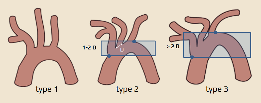
- type 2 and 3 of the aortic arch (based on the vertical distance from the origin of the innominate artery to the top of the aortic arch)
- type 1 (classic) = distance < 1 left CCA diameter
- type 2 = distance is 1-2 CCA diameters
- type 3 = distance is > 2 CCA diameters
- sharp-angle of the CCA
- hypoplasia of the ICA, CCA, or VA

- other variants are less significant
- variant origin of vertebral arteries
- aberrant right subclavian artery (arteria lusoria) (Chrzanowski, 2014)


- instead of being the first branch with the right CCA as the brachiocephalic (innominate) artery, it arises on its own as the fourth branch, distal to the left subclavian artery
- it travels to the right arm, crossing the midline and usually passing behind the esophagus; if the artery compresses the esophagus, it may cause a condition called dysphagia lusoria
- aberrant left subclavian artery (right-sided arch) (Zhyvotovska, 2020)
- an aberrant left subclavian artery arises from the diverticulum of Kommerell, a vascular structure derived from a remnant of the fourth aortic arch
- bronchial arteries (bronchial arteries usually arise from the descending aorta but may arise from the arch)
- common origin of both common carotid arteries

- pseudobovine arch
- a score based on anatomical criteria has been proposed (Snelling, 2018)
- higher scores may predict a longer time from groin puncture to the first pass, lower reperfusion (TICI) scores, and unfavorable clinical outcomes following thrombectomy
Alternative vascular access
Unfavorable vascular anatomy in cervical arteries
- morphological anomalies of the cervical ICA are identified in up to 85% of cases (depending on the age of the population studied and criteria used)
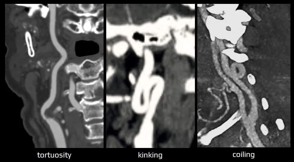
- tortuous ICA ( angle between the CCA and the ICA centerlines is >15° or the course of the ICA is S- or C-shaped)
- kinking (angle between vessel segments is < 90°)
- coiling (circular configuration)
- deformities may be congenital (e.g., fibromuscular dysplasia) or acquired (typically atherosclerosis or age-related loss of elasticity); both leading to vessel elongation and tortuosity
- tortuous cervical anatomy may preclude the optimal positioning of the distal access or intracranial aspiration catheters
- some techniques and newer-generation catheters may be used to overcome cervical vascular tortuosity (Alverne, 2020)
Unfavorable vascular anatomy in intracranial arteries
- severe stenosis with calcifications and the tortuosity of intracerebral arteries may affect the efficacy of endovascular devices (both retrievers and aspiration devices) and is considered an independent predictor of unsuccessful recanalization (Schwaiger, 2015) (Zhu, 2012)
- in highly curved arteries, the stent retriever may lose interaction with the clot, which may escape from the stent (“tapering” phenomenon)
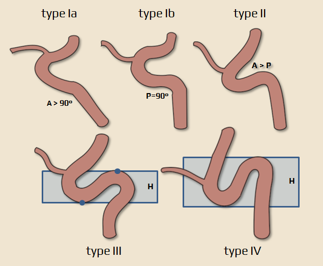
- a four-grade classification system has been developed to identify carotid siphon tortuosity using several features, which consider the angles and height differences of the anterior and posterior genus
- type I – anterior and posterior genu is opened (Ia – posterior genu angle is >90°, type Ib – angle is 90°)
- type II – more acute angle of anterior genu in comparison to type I
- type III – a superior deflection of posterior genu is present
- type IV – the most tortuous type
- ICA tortuosity is measured as a H/(A+P) ratio
- H=height difference of the anterior and posterior genus; A+P=sum of the angles of the anterior (A) and posterior (P) genus
- “saddle” clots positioned at bifurcations
- the clot may extend to both branches of the vessel, and the contact area of the clot with the vessel is extensive
- increased retrieval force is required, but the clot may not be removed or may be pushed to the other branch


