ISCHEMIC STROKE / CLASSIFICATION

SSS-TOAST CLASSIFICATION OF ISCHEMIC STROKE

David Goldemund M.D.
Updated on 05/11/2023, published on 26/09/2022

  • the SSS-TOAST classification is an evidence-based classification algorithm that reflects advancements in stroke imaging and epidemiology [Ay, 2005]
  • based on specific clinical and imaging criteria, each TOAST subtype is further categorized into one of three subcategories: “certain,” “probable,” or “possible.”
  • this new algorithm refines the determination of the most likely etiology, particularly when multiple competing mechanisms are present
  • an automated web-based version of the SSS-TOAST, known as the Causative Classification System (CCS), was developed to facilitate its utilization in multicenter settings (Ay, 2007)
    • CCS enables swift analysis of patient data with excellent intra- and inter-examiner reliability
    • particularly valuable in situations where precise classification is critically important (such as in clinical trials)
    • the text highlighted in purple was not included in the original SSS-TOAST publication in 2005

SSS-TOAST 1 – Large Artery Atherosclerosis (LAA)

Evident
  • occlusion or stenosis ≥50% (according to NASCET) or <50 % diameter reduction with plaque ulceration or thrombosis due to atherosclerosis in clinically relevant extra-/intracranial arteries

AND

  • absence of acute infarcts in vascular territories other than those corresponding to the stenotic or occluded artery
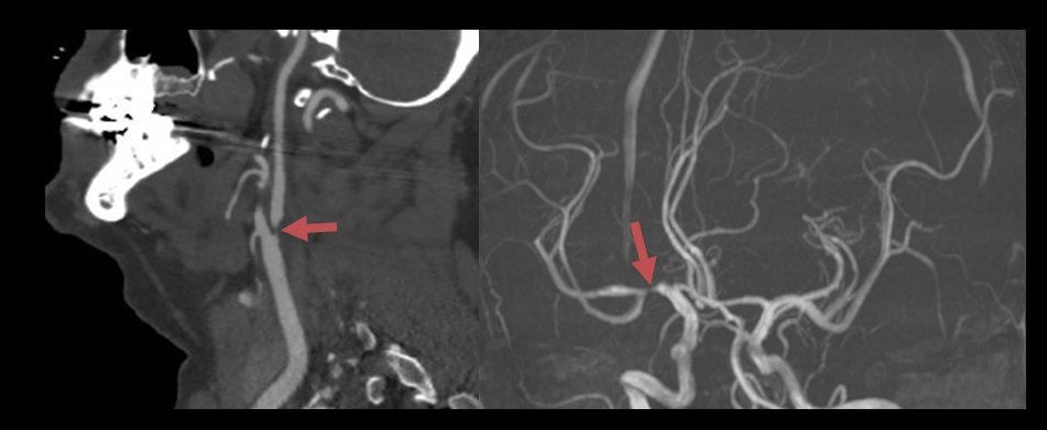
Significant internal carotid artery stenosis on CTA
Large artery atherosclerosis (LAA)
Probable
  • occlusion or stenosis ≥50% (according to NASCET) caused by atherosclerosis in relevant extra-/intracranial arteries
  • history of ≥ 1 TIA/stroke within the territory of the index artery affected by atherosclerosis, occurring within the past month

OR

  • evidence of near-occlusive stenosis or non-chronic total occlusion, determined to result from atherosclerosis, in relevant extra-/intracranial arteries (excluding the vertebral arteries)

OR

  • the presence of ipsilateral border-zone infarctions (BZI) or multiple, temporally separated infarcts occurring within the region supplied by the affected artery


Possible
  • atherosclerotic plaque causing stenosis <50% without any detectable plaque ulceration or thrombosis in a relevant extra-/intracranial artery
  • history of ≥ 2 TIAs/strokes in the territory of index artery affected by atherosclerosis; at least one event occurring within the past month

OR

  • evidence of extensive artery atherosclerosis in the absence of a complete diagnostic workup for alternative causative mechanisms

SSS-TOAST 2 – Cardio-aortic embolism

Evident

The presence of a high-risk cardiac source of cerebral embolism

  • left atrial/ventricular thrombus
  • (paroxysmal) atrial fibrillation/flutter
  • sick sinus syndrome (SSS)
  • recent myocardial infarction (< 1 month)
  • bioprosthetic and mechanical valves
  • rheumatic mitral or aortic valve disease
  • symptomatic congestive heart failure with EF < 30%
  • dilated cardiomyopathy
  • nonbacterial thrombotic endocarditis
  • an intracardiac tumor (such as papillary fibroelastoma or myxoma)
Probable
  • evidence of systemic embolism

OR

  • presence of multiple acute infarcts occurring in close temporal relation within the right and left anterior or both anterior and posterior circulations in the absence of occlusion or near-occlusive stenosis of all relevant vessels
  • other diseases that could potentially lead to multifocal ischemic lesions, such as vasculitides, vasculopathies, and hemostatic or hemodynamic disturbances, are not present
Possible
  • the presence of a cardiac condition associated with low or uncertain risk of cerebral embolism

OR

  • evidence of obvious cardio-aortic embolism in the absence of a complete diagnostic workup for other potential causes

SSS-TOAST 3 – Small-artery occlusion

Evident
  • imaging evidence of a single, clinically relevant acute infarct < 2 cm in greatest diameter within the territory of the penetrating arteries  Lacunar infarct in the left thalamus (DWI)
  • absence of significant pathology in the parent artery at the site of the origin of the penetrating artery (most commonly ostial atheroma, less commonly parent vessel dissection, vasculitis, vasospasm, etc.); parent artery pathology is also referred to as branch artery disease (BAD) / branch occlusive disease (BOD) and is classified as TOAST 1  Penetrating artery disease with a parent artery atherosclerosis

    • diagnosis is based on high-resolution MRI   [Petrone, 2016]
    • more likely in younger patients
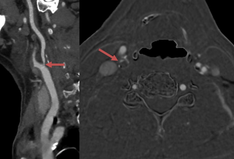
    • infarct is usually larger than a typical arteriolopathic stroke and is typically located in the pons  Probable Branch Artery Disease (BAD) leading to pontine infarct. CTA revealed no major stenosis in posterior circulation. HR-MRI is not available.  [Zhou, 2018]
Probable
  • stereotypic lacunar TIAs within the past week
  • clinical presentation of a classic lacunar syndrome
Possible
  • classical lacunar syndrome in the absence of imaging sensitive enough to detect small infarctions

OR

  • evidence of small artery occlusion in the absence of complete diagnostic assessment for other potential mechanisms

SSS-TOAST 4 – Other causes

Evident
Probable
  • a specific disease process that has occurred in close temporal relation to the stroke onset (e.g., arterial dissection, cardiac or vascular surgery, and cardiovascular procedures
Possible
  • evidence pointing to a different identifiable cause of stroke in the absence of complete diagnostic evaluation for the aforementioned mechanisms

SSS-TOAST 5 – Undetermined causes

  • cryptogenic embolism → ESUS criteria

    • vascular imaging indicative of an embolic origin:
      • abrupt absence of contrast, consistent with a blood clot, within otherwise angiographically normal-appearing intracranial arteries
      • complete recanalization of the previously occluded artery
      • multiple acute infarcts occurring in a close temporal relationship without apparent abnormalities in the relevant arteries
  • other cryptogenic strokes – those not meeting the criteria for cryptogenic embolism
  • incomplete evaluation – the absence of essential diagnostic tests required to determine the underlying cause
  • unclassified – the presence of >1 evident mechanism; either there is probable evidence for each, or no single cause can be reliably established

Related Content

You cannot copy content of this page

Send this to a friend
Hi,
you may find this topic useful:

SSS-TOAST classification
link: https://www.stroke-manual.com/sss-toast-classification/