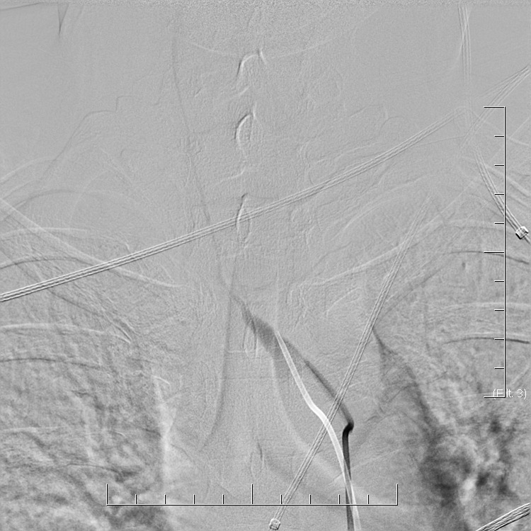
• connective tissue disorders and vasculitides are a heterogeneous group of diseases that may have various manifestations, including stroke
  • vasculitis is characterized by inflammation of the walls of blood vessels and may affect vessels of any size
  • it is a relatively rare cause of stroke (~ <3% of strokes in patients < 50 years of age   [Ferro, 1998]
    • routine testing in an unselected group of patients with ischemic stroke is therefore not useful
  • in cases where the stroke is the initial symptom of the disease, such as in PACNS, diagnosis can be challenging despite extensive investigation, including CSF analysis and brain biopsy
  • vasculitis may cause:
    • stenosis/occlusion
    • development of mural thrombosis with potential distal embolization
    • artery rupture with bleeding (ICH, SAH)
  • probably the most relevant disorders related to stroke neurology are:

Etiopatogenesis

Pathogenetic mechanisms in vasculitides
 I. Immune-mediated
  • type IV immunopathologic reaction
  • autoantibodies
  • immune complexes
    • mostly in secondary vasculitides
    • deposition of immune complexes and C3 complement components can be detected in affected tissues
    • e.g., polyarteritis nodosa, SLE, etc.
 II. Infection, physical or chemical injury (toxins, drugs, medication)
  • infections can indirectly precipitate immune-mediated vasculitis

Classification

  • there are various classifications based on different aspects of the pathological process
  • the following list of vasculitides is not complete; attention is given primarily to those that may affect the cerebral arteries

Classification based on the diameter of the affected vessel

Classification based on the size of most affected vessels
(Names for vasculitides adopted by the 2012 International Chapel Hill Consensus Conference on the Nomenclature of Vasculitides)
Large arteries (aorta and its main branches, including the intracranial ICA segment)
Medium-sized arteries (major organ arteries, arteries forming the circle of Willis)
Small arteries
  • immune complex-induced response
    • cryoglobulinemic vasculitis
    • systemic lupus erythematosus (SLE)
    • IgA vasculitis (Henoch-Schönlein)
    • anti-glomerular basement membrane (anti-GBM) disease (Goodpasture’s disease)
  • the paucity of immune complexes (ANCA-associated vasculitis)
    • eosinophilic granulomatosis with polyangiitis (Churg-Strauss) – (lung, eosinophilia, asthma, p-ANCA)
    • granulomatosis with polyangiitis (Wegener’s) (lung, kidney, positive c-ANCA)
    • microscopic polyangiitis
  • idiopathic, infectious, drugs, chemicals, cancer, and systemic diseases

Primary and secondary vasculitis

 I. Primary vasculitides
II. Secondary vasculitides
  • cancer-associated vasculitis
    • lymphoma
    • malignant histiocytosis
    • neoplastic meningitis
  • infectious vasculitis (proof of infection + vasculitis required)

Clinical presentation

  • general symptoms (often absent in CNS vasculitides)
    • weight loss, fever, fatigue, lack of appetite, increased sweating
    • arthralgia, myalgia
  • nervous system
    • CNS
      • headache
      • epileptic seizures
      • encephalopathy, stroke-like episodes
      • stroke
    • PNS
      • polyneuropathy, mononeuropathy
  • lungs
    • hemoptysis, cough, dyspnoea, asthma, lung infiltrates
  • ENT
    • sinusitis, otitis, deafness
  • skin:
    • purpura, pyoderma, necrosis
  • eye:
    • uveitis, iridocyclitis, episcleritis
  • kidneys:
    • renal hypertense
    • proteinuria
    • erythrocyturia
    • renal failure
Course Signs and symptoms of CNS involvement
 acute/subacute
subacute

relapsing-remitting

headache
epileptic seizures
stroke-like episodes
ischemic stroke, IC bleeding
encephalopathy
extrapyramidal disorders (chorea, myoclonus)
optic nerve disorders
cranial neuropathies

Diagnostic evaluation

In general, the diagnosis of CNS vasculitis is very rare (< 1-3%), but also extremely difficult (especially with PACNS). It occurs predominantly in younger patients (except for temporal arteritis) without traditional vascular risk factors, manifestations/history of systemic disease, typical clinical course, and imaging findings (parenchymal and arteries involvement). Arteries may show variable degrees of stenosis, occlusion, and contrast enhancement in the wall. The correlation of neuroimaging with clinical presentation and laboratory tests helps establish the diagnosis.

  • only a positive biopsy can provide a definitive diagnosis of CNS vasculitis
  • if the biopsy is not performed or is negative (may be false negative), then in the presence of suspicious clinical and radiologic findings, possible CNS vasculitis can be discussed
    • clinical findings indicative of vasculitis: headache (70%) and encephalopathy (up to 95%) or seizures
    • no specific brain imaging pattern
    • vascular imaging is typical in Takayasu arteritis and temporal arteritis
Laboratory tests
  • basic inflammatory parameters
    • ↑CRP
    • ↑ESR
  • blood tests for autoimmune diseases
    • complement complex (lupus)
    • p-ANCA, c-ANCA
      • granulomatosis with polyangiitis, eosinophilic granulomatosis with polyangiitis, inflammatory bowel disease
    • anti-dsDNA (SLE)
    • ANA antibodies (SLE)
      • most commonly used to diagnose lupus; these antibodies can sometimes also signal other systemic autoimmune diseases such as rheumatoid arthritis, scleroderma, or Sjögren’s syndrome
    • ENA panel (Extractable Nuclear Antigen)
      • anti-Sm
      • anti-RNP
      • anti-SSA, anti- SSB (Sjögren Syndrome)
      • ani-Scl-70 (scleroderma)
      • anti-Jo-1
    • antiphospholipid antibodies (SLE)
      • LA
      • ACLA
      • anti-β2-glycoprotein I (anti-β2GPI)
    • RF (rheumatoid factor)
      • used to diagnose rheumatoid arthritis
      • normal ranges: < 15 IU/mL, < 1:80 for titer levels
  • routine blood tests
    • basic metabolic panel
      • to assess problems with the pancreas, liver, heart, or kidneys
    • complete blood count (CBC)
      • normochromic anemia, leukocytosis, eosinophilia
    • coagulation studies
  • CSF examination
    • abnormal in up to 95%
    • pleocytosis (less commonly mild mononuclear pleocytosis)
    • proteinorhachia (~50-80%)
    • oligoclonal bands (OCB); positive in (~50% of cases
    • consider testing for autoimmune and paraneoplastic encephalitis antibodies
  • basic infectious disease screening (Lyme disease, herpes viruses, HIV, syphilis)

Normal ESR ranges

The normal ranges for ESR are:

  • 0-15 mm/h for men < 50
  • 0-20 mm/h for men > 50
  • 0-20 mm/h for women <50
  • 0-30 mm/h for women > 50
  • 0-10 mm/h for children
  • 0-2 mm/h for infants

CRP ranges

  • < 0.3 mg/dL – normal, which is the level for most healthy adults
  • 0.3-1 mg/dL – normal or mild elevation can be seen with obesity, pregnancy, depression, diabetes, common cold, gingivitis, periodontitis, sedentary lifestyle, smoking, and genetic polymorphisms
  • 1-10 mg/dL – moderate elevation indicates systemic inflammation, such as in the case of rheumatoid arthritis, systemic lupus erythematosus (SLE), or other autoimmune diseases, malignancy, myocardial infarction, pancreatitis, and bronchitis
  • > 10.0 mg/dL – marked elevation signals acute bacterial infection, viral infection, systemic vasculitis, and major trauma
  • > 50.0 mg/dL -severe elevation may be caused by acute bacterial infections
Brain imaging
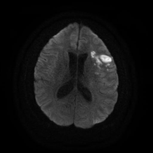
  • MRI is more advantageous than CT
  • changes variable and mostly nonspecific
    • cortical and subcortical T2 hyperintense lesions + periventricular white matter lesions Wegener's granulomatosis with non-specific white matter hyperintensities (FLAIR)
    • sometimes, postcontrast enhancement of the lesions or dural enhancement is present
    • MRI may show associated lesions in the ENT region (e.g., in GPAT1 postcontrast dural enhancement in Wegener's granulomatosis [Allen, 2007]
  • MRI may be completely negative even in the presence of vasculitis [Alhalabi, 1994]
Vascular imaging

Angiography

  • CTA/MRA/DSA)
    • CTA performed from the aortic arch is considered the baseline imaging technique; ultrasound may be used to diagnose and monitor stenosis dynamics (or to differentiate spasm and RCVS)
    • exclude atherosclerotic involvement in typical sites (artery bifurcation, carotid siphon, M1 segment of the MCA); calcifications are common in these cases
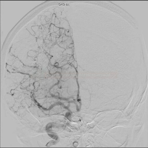
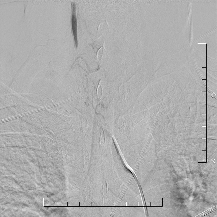
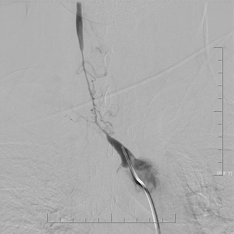
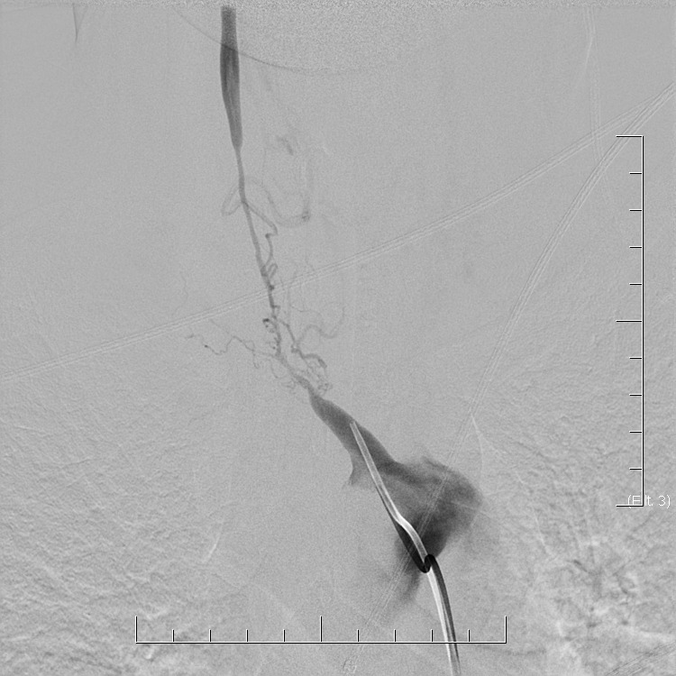
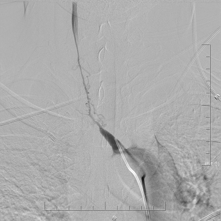
    • vasculitis is characterized by segmental, multifocal involvement, alternating narrowed and dilated segments, without accompanying calcification (CAVE combination of vasculitis in a patient with atherosclerotic involvement) CTA and DSA findings of multiple stenoses in primary cerebral vasculitis with fulminant course
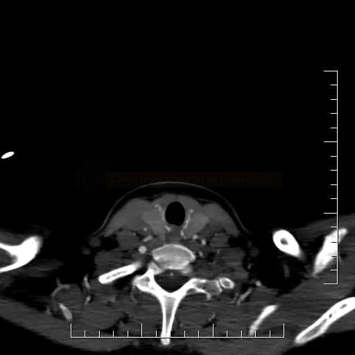
    • Takayasu arteritis is characterized by concentric thickening of the aortic wall and outlying branches Takayasu arteritis on CTA - typical concentric enlargement of the wall in affected arteries  Takayasu arteritis on CTA
    • vasculitis may have negative angiographic findings due to the involvement of small arteries  (20-40% of cases)
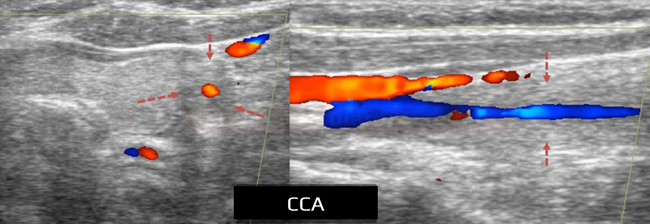
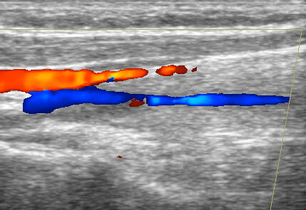
  • neurosonology
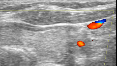
    • detection of intracranial stenoses on Doppler (nonspecific finding)
    • concentric wall thickening in extracranial arteries  

Vessel wall imaging

  • post-contrast high-resolution dark (black) blood MRI sequences – “wall imaging”
  • wall imaging must be assessed with caution
    • enhancement does not always reflect an underlying vasculopathy or vasculitic origin (Kang, 2021)
    • false-positive findings occur:
      • due to poststenotic low flow
      • after mechanical recanalization
      • due to leptomeningeal enhancement or enhancement of organized thrombus
PACNS

Takayasu on CTA

Biopsy
  • except for PACNS, biopsy should preferably be performed in the periphery (skin, muscle, nerve, kidney, superficial temporal artery, etc.)
  • brain biopsy should be taken from the affected area of the non-dominant hemisphere or the temporal lobe pole of the non-dominant hemisphere
    • leptomeninges should be part of the sample (higher detection of vasculitic changes in meningeal vessels)
    • open biopsy is more productive than stereotactically guided biopsy  [Torres, 2016]
  • positive biopsy confirms a “definitive” diagnosis of CNS vasculitis (in the absence of a biopsy or with a negative biopsy, we can speak of “possible” CNS vasculitis)
    • inconsistent results  between angiographic and biopsy findings are relatively common, and false negative biopsies may occur (in 25-40%)   [McVerry, 2017]
    • a positive biopsy is found in about 9-36% of cases
  • serious complications occur in < 4% of cases  [Torres, 2016]
  • many presumed “vasculitides” have a different etiology according to biopsy:
    • one study found vasculitis in 36%, negative findings in 25%, and other etiologies (lymphoma, SM, infection) in 39% [Alrawi, 1999]
    • according to another paper, PACNS was found in only 11% of suspected patients, and another etiology was found in 30% (encephalitis, lymphoma, CAA)  [Torres, 2016]
Other examinations
  • X-ray (lungs, paranasal sinuses, etc.)
  • CT of abdomen + chest (malignancy, exclusion of sarcoidosis, etc.)
  • EMG – evidence of neuropathy
  • ophthalmologic examination
  • FDG PET/CT

Management

Detailed management and dosing is discussed in individual vasculitides

Remission induction

  • corticosteroids (IV bolus)
  • immunosuppressive drugs
    • pulse cyclophosphamide
    • cyclophosphamide PO
  • plasmapheresis
  • intravenous immunoglobulins (IVIG)
  • monoclonal antibodies
    • rituximab

Remission maintaining

  • PO corticosteroids + immunosuppressive drugs
    • azathioprine
    • methotrexate
    • mycophenolate
    • cyclophosphamide

Related Content

You cannot copy content of this page

Send this to a friend
Hi,
you may find this topic useful:

Vasculitis
link: https://www.stroke-manual.com/vasculitis/