ISCHEMIC STROKE / CLASSIFICATION AND ETIOPATHOGENESIS
Fabry disease
Updated on 26/12/2023, published on 10/05/2023
Etiopathogenesis
- Fabry disease (FD) is a rare inherited lysosomal storage disorder that can cause severe damage in multiple organs, particularly the kidneys and heart
- first described by Anderson and Fabry in 1898 as angiokeratoma corporis diffusum (also known as Anderson-Fabry disease)
- FD is caused by mutations in the α-galactosidase A (α-Gal-A) gene located on the X chromosome

- more than 400 known mutations of the GLA gene cause a complete or partial defect in the lysosomal enzyme alpha-galactosidase (α-Gal-A), which is responsible for the cleavage of glycosphingolipids
- the primary substrate, globotriaosylceramide (Gb3), cannot be degraded by other pathways and accumulates in lysosomes, affecting cellular functions and causing organ complications
- women have a 50% chance of passing the gene on to their children
- men with Fabry disease pass the defective gene to all their daughters, while sons are healthy
- cells mainly affected:
- vascular endothelium and vascular smooth muscle cells
- kidney cells
- myocardial cells
- nerve cells
- phenotype varies depending on the specific gene defect and residual enzyme activity; the severe form may be seen in both males and heterozygous females
- male patients
- disability is more pronounced and manifests earlier (males are hemizygous, and the disease has a high penetrance ⇒ classic phenotype)
- late-onset forms have also been reported, depending on the degree of individual enzymatic impairment
- female patients
- variable expression (asymptomatic to severe) due to the random inactivation of one of the X chromosomes (the female body is thus a mosaic of cells in which either the healthy or the diseased X chromosome is active)
- unlike other X-linked diseases such as hemophilia, the population of cells expressing the healthy chromosome is unable to correct the metabolic defect in cells where the chromosome carrying the gene mutation is active
- male patients
- the disease can become severe and fatal with advanced brain, renal, or cardiac involvement
- early diagnosis and enzyme replacement therapy (ERT) reduce morbidity and mortality
- without treatment, patients typically do not survive beyond the age of 40
Stroke is most commonly caused by:
- dolichoectasia
- cardioembolism
- arteriolopathy
Clinical presentation
Neurologic symptoms
- acroparesthesia – pain, tingling, and burning in the hands and feet – often one of the first symptoms
- mostly neuropathic pain due to the involvement of nociceptive nerve fibers provoked by heat, exertion, and stress
- autonomic dysfunction
- GIT disorders associated with impaired bowel motility (frequent diarrhea, feeling of early satiety)
- decreased heart rate variability
- hypo-/anhidrosis (from damage to sweat cells and the autonomic nervous system)
- recurrent headaches
- vestibulocochlear system impairment
- vertigo and hearing loss
- vascular disease (usually manifesting in the 3rd decade of life)
- stroke (prevalence of Fabry disease in young men with cryptogenic stroke is reported to be < 1%)
- lacunar infarcts
- infarcts with a predilection for the posterior circulation due to dolichoectasia
- infarcts attributed to cardioembolism
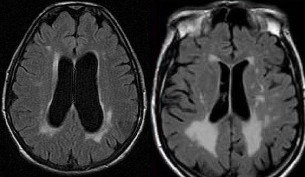
- ischemic leukoencephalopathy due to a progressive arteriolopathy (initially asymptomatic, but later may lead to vascular cognitive impairment)
- stroke (prevalence of Fabry disease in young men with cryptogenic stroke is reported to be < 1%)
Renal symptoms
- from the 2nd decade, a progressive course occurs due to glomerular and tubular cell damage
- microalbuminuria appears first, followed by proteinuria
- especially in males, the disease progresses with a gradual decline in glomerular filtration to renal failure with the need for dialysis therapy or kidney transplantation, typically in the 4th decade
- the fate of the transplanted kidney in Fabry disease is generally good
- the diagnosis is often made by a renal biopsy performed for the evaluation of proteinuria
- with conventional fixation of the biopsy material, lipid inclusions in the cells may not be visible
- the classic electron microscopic image shows zebrafish-like bodies of accumulated Gb3
- with conventional fixation of the biopsy material, lipid inclusions in the cells may not be visible
Cardiologic disorders
- cardiac involvement is very complex – pathological changes are evident not only in endothelial cells of arterioles and capillaries but also in smooth muscle cells
- reduced coronary reserve, endothelial dysfunction, cardiomyopathy, and myocardial fibrosis ⇒ cardiac failure
- arrhythmias
- conduction disturbances in the form of fascicular block or AV block of varying degrees
- these patients require pacemaker implantation
- atrial flutter and fibrillation ⇒ cardioembolism
Skin and eye symptoms
- angiokeratoma (Resende de Jesus, 2018)


- papular red spots, ~ 1 mm in size, protruding above the surface
- preferentially located in the lower abdomen, genitalia, buttocks, flanks, and extensor surfaces of the upper limbs, less frequently on mucous membranes and other parts of the body
- onset typically in childhood, progressive over time
- angiectasia – small, slightly raised, purple-red dilated blood vessels. The number and size of the lesions increase with age
- lymphedema
- cornea verticillata – the corneal hyperdensities


- can be observed using a slit lamp as whitish streaks
Diagnostic evaluation
Clinical presentation
- “cryptogenic” stroke in young age + nephropathy + neuropathy with acroparesthesia + cardiopathy + skin (angiokeratoma) and eye lesions (cornea verticillata)
Imaging methods
- brain MRI
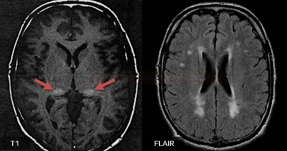
- leukoencephalopathy (white matter lesions) with T2 hyperintensities (nonspecific for Fabry disease)
- pulvinar sign – increased signal on T1-weighted images [Takanashi, 2003]
- thought to result from the deposition of glycolipids or calcium) [Cocozza, 2018]
- its absence does not rule out the disease (low specificity and sensitivity)
- lacunar infarcts
- microbleeds on GRE
- some MRI changes may resolve with early specific treatment
- vascular imaging (MRA, CTA)
- TTE
- renal imaging
- nonspecific findings such as increased echogenicity and thinned renal cortex, as well as multiple renal cysts (typically small, uniform in size, located beneath the capsule)
- the pulvinar nucleus is the largest nucleus of the thalamus and plays a role in visual attention and modification of behavior response
- the PN accounts for 30% of the volume of the thalamus and is supplied by the posterior choroidal artery
- the pulvinar sign is defined as a bilateral, symmetrical pulvinar high signal relative to the signal intensity of other deep grey matter nuclei and cortical grey matter on unenhanced T1-weighted brain, FLAIR, or DWI MRI imaging
- various substances increase the intensity of T1 imaging such as:
- fat
- MRI fat suppression may confirm diagnosis
- MRI fat suppression may confirm diagnosis
- calcium (calcium deposition presents as hyperdensity on CT scans)
- at lower calcium concentrations below 30–40%, hyperintensity is seen on T1 weighted images. However, when there is an increasing concentration above 30–40%, the hyperintensity disappears (Moore, 2003)
- iron
- manganese
- melanin
- fat
- pulvinar sign may be seen in Fabry disease but is not pathognomic
- incidence ~ 23% (over 30% by age 50 years) (Moore, 2003)
- low specificity and sensitivity
- probably caused by calcifications
- the lack of signal intensity change when using MR fat suppression excluded fat as a possible agent in one study (Cocozza, 2018)
- incidence ~ 23% (over 30% by age 50 years) (Moore, 2003)
- the pulvinar sign has been described in numerous neurological disorders
- Tay Sachs, Krabbe disease, CNS infections, radiation, variant Creutzfeldt-Jakob disease, anti-CV2 encephalitis, Wernicke´s encephalopathy, anti-HU encephalitis, limbic encephalitis, neurosarcoidosis, and post-iInfluenza encephalitis
Laboratory test
| Content available only for logged-in subscribers (registration will be available soon) |
Management
Specific therapy
Symptomatic therapy
- stroke prevention – antiplatelets/anticoagulants
- treatment of pain and acroparesthesia (phenytoin, carbamazepine, gabapentin, pregabalin)
- nephroprotective treatment
- ACE-I, AT receptor blockers (sartans)
- spironolactone
- treatment of GIT disorders
- prokinetics
- pancreatic lipase
- H2 blockers
- cardiological treatment
- antiarrhythmic drugs, cardioverter implantation
- management of CAD
- treatment of atrial fibrillation
heart transplantation
- additional measures to minimize triggers of painful attacks (prompt treatment of fever and infections, maintaining adequate physical activity, using air conditioning, ensuring adequate hydration, etc.)
FAQs
- Fabry disease is a rare genetic disorder caused by mutations in the enzyme alpha-galactosidase A (GLA) gene
- the deficiency of GLA leads to the accumulation of glycolipids in various body tissues, affecting organs and causing a range of symptoms
- Fabry disease follows an X-linked recessive inheritance pattern; affected individuals have mutations on one of their X chromosomes
- since males have only one X chromosome, they are more severely affected
- females can be carriers and may exhibit symptoms
- common symptoms include pain in the hands and feet (acroparesthesia), skin rashes (angiokeratomas), gastrointestinal issues, corneal opacities, hearing loss, and organ involvement such as kidney and heart problems.
- diagnosis often involves genetic testing to identify mutations in the GLA gene, enzyme assays to measure alpha-galactosidase A activity, lipid analysis, and clinical evaluation
- enzyme replacement therapy (ERT) is one option to replace the missing enzyme
- chaperone therapy and supportive care for specific symptoms are also used
- kidney transplantation may be necessary in cases of severe renal impairment
- long-term complications can include kidney failure, heart disease, stroke, hearing loss, and reduced quality of life. Early diagnosis and appropriate treatment can help mitigate these complications
- the prognosis varies depending on the severity of symptoms and the timing of diagnosis and treatment
- early diagnosis and appropriate management can improve the long-term outlook and quality of life
- Fabry Disease is a rare disorder, estimated to affect approximately 1 in 40,000 to 60,000 males
- female carriers may also exhibit symptoms


