INTRACEREBRAL HEMORRHAGE / VASCULAR MALFORMATIONS
Cerebral venous angioma (DVA)
Updated on 24/06/2024, published on 20/09/2023
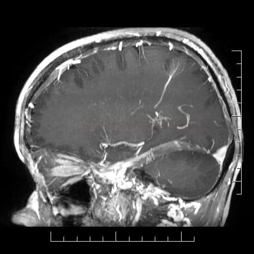
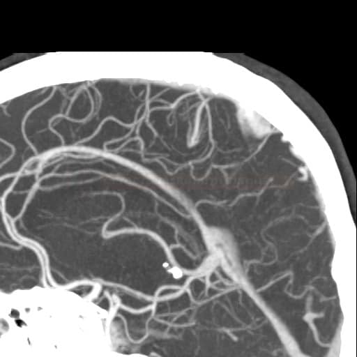
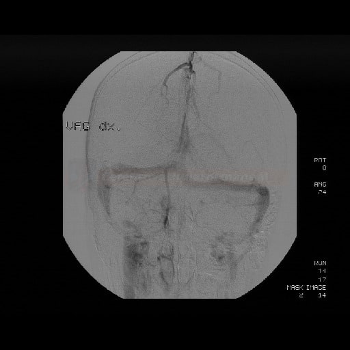
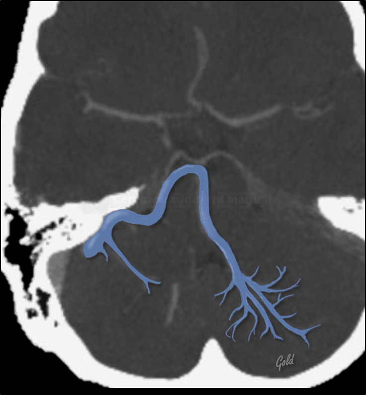
- cerebral venous angioma, also known as Developmental Venous Anomaly (DVA), is a congenital malformation where dilated veins with no abnormal feeding artery (caput medusae sign) converge into a single large abnormal draining vein
- DVAs rarely bleed ⇒ if a hemorrhage is present, look for associated cavernous malformation (15-20%), AVM, or a tumor
- most commonly, DVAs are localized in the parietal and frontal lobes (up to 64%) and cerebellum (up to 30%)
- supratentorial drainage ⇒ superficial or deep subependymal veins
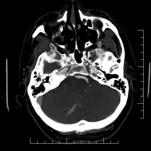
- infratentorial drainage ⇒ the transverse sinus, the great vein of Galeni, petrous sinus → Anatomy of cerebral veins and sinuses
Clinical presentation
- commonly asymptomatic (usually an incidental finding on neuroimaging)
- epileptic seizures
- headaches
- venous infarction (in case of rare thrombosis of the draining vein)
- intracerebral bleeding is rare (0.2-0.4% per year)
- increased risk is associated with occlusion of the draining vein, e.g., due to thrombosis [Agarwal, 2014]
- search for an associated cavernous malformation (cavernoma)
Diagnostic evaluation
Differential diagnosis
- sometimes, there is an atypical imaging appearance, which may be confounded by concurrent pathology
- dural arteriovenous fistula (DAVF)
- arteriovenous malformation (AVM)
- dural sinus thrombosis
- Sturge-Weber syndrome
- DVA may mimic thrombosed cerebral vein (Althobaiti, 2019)
Management
- a conservative approach is common for isolated DVA
- the draining vein often serves as a drainage for the surrounding unaffected areas
- surgical occlusion or spontaneous thrombosis may lead to venous infarction
- if needed, treat concurrent malformations
FAQs
- a benign cerebral vascular malformation in which dilated veins converge into a single large draining vein
- there is no abnormal feeding artery
- adjacent brain parenchyma is normal
- DVAs are considered congenital
- DVA is primarily diagnosed by MRI or CT angiography
- SWI is the preferred MRI sequence to detect venous malformations
- generally asymptomatic (typically incidental finding on neuroimaging)
- symptoms, if present, are usually secondary to associated lesions (CCM, AVM)
- thrombosis of the draining vein is an extremely rare complication leading to venous infarction
- AVM has large feeding arteries, tortuous vessels, and abnormal adjacent brain parenchyma that are not observed in DVAs
- seizures may occur in the presence of an associated epileptogenic lesion (e.g., cavernoma or dysplasia) or as a result of complications (e.g., venous infarction)
- typically, no intervention is needed unless associated with other vascular malformations (AVM, cavernous malformation) or symptomatic hemorrhage
- surgical intervention is rarely indicated and generally not recommended due to the role DVAs play in venous drainage
- excellent, particularly when isolated; there is no increased risk of bleeding in isolated DVAs
- typically, routine follow-up is not required unless DVA is associated with other vascular anomalies

