ADD-ONS / ANATOMY
Anatomy of cerebral veins and sinuses
Updated on 16/07/2024, published on 09/04/2021
Classification
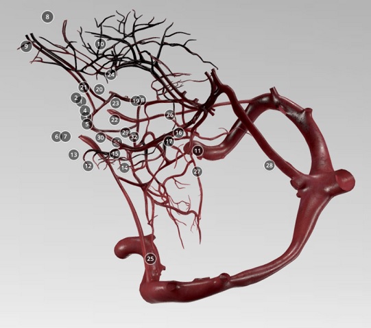
- venous blood is drained from the brain by a system of cerebral veins (vv. cerebri), which are emptied into dural sinuses and cervical veins (internal jugular veins and vertebral veins)
| Cerebral veins |
|
| Dural venous sinuses (dural sinuses, cerebral sinuses) |
|
| Meningeal veins |
|
| Ophthalmic veins |
|
| Diploic veins |
|
| Emissary veins |
|
| Veins of labyrinth |
The cerebral veins
- cerebral veins drain blood from the brain parenchyma to the dural venous sinuses
- veins are characterized by a larger caliber than the cerebral arteries, have thin walls, and lack muscular tissue and valves
- typically, they have a variable course and numerous anastomoses
- cerebral veins can be divided into:
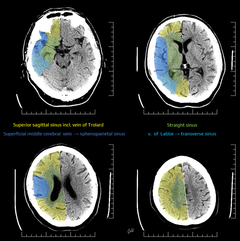
- superficial (cortical) cerebral veins → draining to the superior sagittal sinus (sinus sagittalis superior)
- deep (subependymal) cerebral veins → draining to the straight sinus (sinus rectus)
- superficial (cortical) cerebral veins → draining to the superior sagittal sinus (sinus sagittalis superior)
Superficial (cortical) cerebral veins
- superior cerebral veins
- collect blood from the brain convexity and medial surfaces and drain into the superior sagittal sinus
- middle cerebral veins – the most important is the superficial middle cerebral vein (Sylvian vein)
- usually courses along the Sylvian fissure and receives numerous small tributaries from the opercular areas. It curves anteriorly and empties either into the sphenoparietal sinus or directly into the cavernous sinus
- has anastomoses with:
- the deep middle cerebral vein
- the superior sagittal sinus (superior anastomotic vein – vein of Trolard). This vein is usually smaller than the superficial middle cerebral vein and vein of Labbé
- the anterolateral portion of the transverse sinus (inferior anastomotic vein – vein of Labbé)
- inferior cerebral veins
- collect blood from the basal parts of the frontal, temporal, and occipital lobes
- drain into the superficial middle cerebral vein, cavernous sinus, superior petrosal sinus, transverse sinus, and some drain into the inferior sagittal sinus
Deep cerebral veins
- deep veins and their tributaries drain the thalamus, hypothalamus, corpus striatum, internal capsule, corpus callosum, septum pellucidum, choroid plexus, and the white matter of the cerebral hemispheres
- medullary veins: numerous veins traverse the deep medullary white matter, draining into subependymal veins
- subependymal veins (receive medullary veins and aggregate into greater vessels)
- deep middle cerebral veins
- arises from the depth of the lateral fossa and enters the basal vein of Rosenthal
- anastomoses with the superficial middle cerebral vein
- its tributaries include, among others, inferior thalamostriatal veins and the choroidal vein (draining the choroid plexus)
- arises from the depth of the lateral fossa and enters the basal vein of Rosenthal
- the basal veins of Rosenthal
- paired veins originating near the anterior perforated substance and running posteriorly and medially
- each of these veins is formed by the union of the anterior cerebral vein, deep middle cerebral vein, and some inferior striate veins
- they drain into the vein of Galen together with internal cerebral veins
- they have mutual anastomosis
- internal cerebral veins
- formed close to the interventricular foramen (of Monro) by the confluence of the choroidal vein, the thalamostriate vein, and the veins of the septum pellucidum (they usually join via the thalamostriate vein)
- the drainage area is variable and usually includes the thalami and periventricular white matter
- the internal cerebral veins, together with the basal veins of Rosenthal, merge into the great cerebral vein (of Galen)
- the great cerebral vein (of Galen) with inferior sagittal sinus together form the straight sinus
- deep middle cerebral veins


Other intracranial veins
- meningeal veins
- meningeal veins collect blood from the meninges and the inner surfaces of the cranial bones
- they drain into dural venous sinuses, most notably the superior sagittal sinus, the transverse sinuses
- meningeal veins are significant in the context of cranial injuries where dural penetration can lead to venous bleeding.
- diploic veins
- large, valveless veins with a thin vascular wall situated within the diploe between the inner and outer layers of the cranial bones
- drain blood from the diploe to either the dural venous sinuses or the scalp veins
- veins are connected with the cerebral sinuses via emissary veins
- subtypes include frontal diploic vein, anterior temporal diploic vein, posterior temporal diploic vein, occipital diploic vein
- emissary (skull) veins (vena emissaria)
- frontal, condylar, mastoid, occipital, and parietal emissary veins pass through foramina in the skull to provide a venous communication between the dural venous sinuses and veins of the scalp or veins inferior to the skull base (cranial-cerebral anastomosis)
- both diploic and emissary veins provide venous drainage across the skull and can serve as conduits for infection or metastases
- ophthalmic veins
- ophthalmic veins (superior, inferior) connect the facial and angular veins to the cavernous sinus
- inferior vein may also communicate with the pterygoid venous plexus through the inferior orbital fissure
- ophthalmic veins are important in the spread of infections from the face (danger triangle of the face) and may also be involved in thrombotic conditions affecting the cavernous sinus
- prominent on imaging when dilated, which may indicate carotid-cavernous fistula, thrombosis, or other orbital pathology
- ophthalmic veins (superior, inferior) connect the facial and angular veins to the cavernous sinus
- cerebellar veins
- superior cerebellar veins are formed by the union of the precentral cerebellar vein and superior vermian vein. They collect blood from the upper surface of the cerebellum and drain into the great vein (or into the straight, transverse sinus or superior petrosal venous sinuses).
- inferior cerebellar veins collect blood from the inferior surface of the cerebellum and drain into the transverse, occipital, or sigmoid sinuses
- brainstem veins
- brainstem veins drain the medulla, pons, and mesencephalon, including the cerebral peduncles, tegmentum, and quadrigeminal plate
- there are multiple connections into the inferior and superior petrosal sinuses, to the basal vein of Rosenthal, and caudally to spinal veins
- some of the brainstem veins:
- peduncular veins
- lateral mesencephalic veins
- median or lateral anterior pontomesencephalic vein(s)
- transverse pontine veins
- median or lateral anterior medullary vein(s)
- transverse medullary veins
Dural venous sinuses
- dural venous sinuses are channels located between the two layers of the dura mater that form the main drainage pathways of the brain, predominantly to the internal jugular veins (IJV)
- they run alone, not parallel to the arteries
- they are valveless, without a muscular layer in the wall ⇒ no flow regulation, possible bidirectional blood flow
- they receive:
- blood from the cerebral veins
- cerebrospinal fluid (CSF) from the subarachnoid space (via arachnoid granulations)
| Unpaired | Paired |
| superior sagittal sinus inferior sagittal sinus straight sinus occipital sinus intercavernous sinus |
transverse sinus sigmoid sinus superior petrosal sinus inferior petrosal sinus cavernous sinus sphenoparietal sinus basilar venous plexus |
- superior sagittal sinus (SSS)
- the largest dural venous sinus, which runs along the superior aspect of the falx cerebri (from the foramen caecum anteriorly to its termination at the confluence of sinuses)
- receives venous blood from numerous superficial cortical veins and is connected with the superficial middle cerebral vein (via the vein of Trolard)
- there are frequent anatomic variations
- hypoplasia of the middle part
- variations in the anterior (rostral) segment
- duplication of the rostral portion
- duplication with unilateral hypoplasia
- complete or bilateral hypoplasia of duplicated sinus
- inferior sagittal sinus (ISS)
- runs along the inferior edge of the falx cerebri and drains blood into the straight sinus (merges with the great vein of Galen)
- It receives tributaries from the falx itself as well as some small veins from the medial surface of the hemispheres
- straight sinus
- located at the junction between the falx cerebri and the tentorium cerebelli
- it receives blood from the inferior sagittal sinus and the vein of Galen, and some of the superior cerebellar veins
- runs posteroinferiorly towards the confluence of sinuses (approx. in 60% of cases)
- sometimes, it terminates in the left or right transverse sinus
- may be duplicated or hypoplastic (if hypoplastic or absent, a persistent falcine sinus is present, draining blood directly into the SSS)
- located at the junction between the falx cerebri and the tentorium cerebelli
- occipital sinus
- lies on the inner surface of the occipital bone, receiving tributaries from the marginal sinus of the foramen magnum (which is connected to the vertebral venous plexus)
- sinus ends in the confluence of the sinuses
- may be quite extensive and off-midline (his position must be verified before surgery in the posterior fossa)
- transverse sinus
- paired sinus arising from the confluence of the superior sagittal, occipital, and straight sinuses (confluence of sinuses)
- runs along the lateral border of the tentorium cerebelli and ends in the sigmoid sinus, just as it receives the superior petrosal sinus
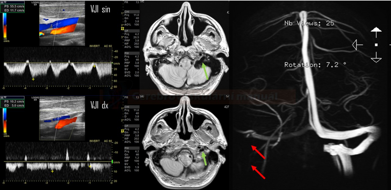
- transverse sinuses are usually asymmetrical (hypo- or aplasia of the left sinus occurs in almost 60% of cases)
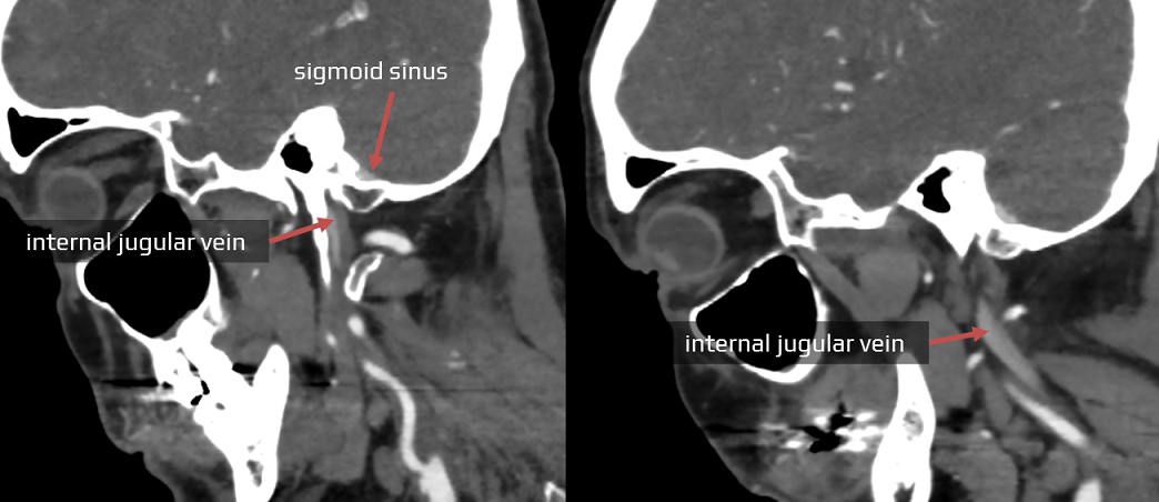
- sigmoid sinus
- typically continues from the transverse sinus
- passes inferiorly in an “S”-shape and heads toward the jugular foramen, ending in the jugular bulb; from here, it continues as the internal jugular vein (IJV)
- the sigmoid sinus is at risk during surgical procedures involving the mastoid process such as mastoidectomy
- superior petrosal sinus
- a narrow dural venous sinus located within the anterolateral margin of the tentorium cerebelli. It spans from the cavernous to the transverse sinus
- its function is to drain the venous blood from the brainstem, temporal lobe, cerebellum, middle and inner ear into the transverse sinus
- It also connects with the inferior petrosal sinuses and the basilar plexus
- inferior petrosal sinus
- a paired cranial venous channel that drains the cavernous sinus, midbrain, cerebellum, and inner ear
- emerges from the cavernous sinus within the middle cranial fossa and empties into the internal jugular vein
- may also drain into the suboccipital external vertebral venous plexus
- cavernous sinus (CS)
- a paired sinus on either side of the pituitary fossa and body of the sphenoid bone, spanning from the orbit apex to the apex of the petrous temporal bone
- divided by numerous fibrous septi into a series of small caverns
- connected anteriorly and posteriorly by transverse junctions (intercavernous sinus), forming a closed venous ring
- the lateral wall of the CS is penetrated by the oculomotor, trochlear, and ophthalmic nerves. The center of the sinus is penetrated by the terminal segment of the ICA and the abducent nerve
- receives venous blood from:
- inferior and superior ophthalmic veins (coming from the orbit through the superior orbital fissure. Via this route, the CS is connected with the veins of the face (pterygoid plexus – facial and angular vein – ophthalmic vein – CS), allowing for potential intracranial spread of inflammation
- sphenoparietal sinus
- superficial middle cerebral vein
- inferior and superior ophthalmic veins (coming from the orbit through the superior orbital fissure. Via this route, the CS is connected with the veins of the face (pterygoid plexus – facial and angular vein – ophthalmic vein – CS), allowing for potential intracranial spread of inflammation
- drainage is via:
- superior petrosal sinus → the transverse sinus
- inferior petrosal sinus → the jugular bulb
- venous plexus on the internal carotid artery (ICA) → the basilar venous plexus
- sphenoparietal sinus
- located along the posteroinferior ridge of the lesser wing of the sphenoid bone
- drains into the CS and receives tributaries from the superficial middle cerebral vein and middle meningeal vein
- basilar venous plexus
- lies on the inner surface of the clivus and connects numerous regional venous structures:
- superiorly – cavernous sinuses, superior petrosal sinuses, intercavernous sinuses
- laterally – inferior petrosal sinuses
- anteriorly – clival diploic veins
- inferiorly through the foramen magnum – vertebral venous plexus, marginal sinus
- lies on the inner surface of the clivus and connects numerous regional venous structures:
Main cervical veins
- there are three main jugular veins in the neck (external, internal, and anterior)
- they are ultimately responsible for the venous drainage of the entire head and neck
- in the supine position, blood from the head is primarily drained via the internal jugular veins (IJV)
- in the upright position, the IJV collapses (due to a drop in hydrostatic pressure), and drainage is mediated predominantly by the vertebral veins
- the cross-section area and shape of the IJV varies with head and body position
Internal jugular vein (IJV)
- a paired vessel located within the carotid sheath on each side of the neck
- drains venous blood from most of the skull, brain, and superficial structures of the head and neck
- originates within the jugular foramen as a continuation of the sigmoid sinus; its origin is determined by a dilation known as the superior bulb
- descends in the neurovascular bundle of the neck (lateral to the ICA and CCA, and the vagus nerve )
- before its termination, it has another dilation (inferior bulb), sometimes with a valve above it
; the morphology of the valve varies:
- VJI terminates posterior to the sternal end of the clavicle, merging with the ipsilateral subclavian vein to form the brachiocephalic (innominate) vein
Vertebral vein
- a paired vessel located in the transverse foramina of the cervical vertebrae on either side of the neck
- originates from the suboccipital plexus and traverses the transverse foramina of the C1-6, surrounding the vertebral artery (this plexus is considered the proximal part of the vertebral vein)
- at the level of the C6 transverse foramen, the plexus merges into a single vessel
- the vertebral vein then courses inferiorly and empties into the brachiocephalic (innominate) vein via the orifice, which usually has a bicuspid valve
- brachiocephalic veins merge to form superior vena cava


