ADD-ONS / ANATOMY
Blood Supply of the Spinal Cord
Updated on 18/11/2023, published on 04/03/2022
The arterial system
Spinal arteries
- the spinal arteries originate primarily from the vertebral arteries (VAs) and run longitudinally from the medulla oblongata to the medullary cone (conus medullaris)
- the VAs serve as the primary blood supply to the upper spinal cord
- spinal arteries receive reinforcement from segmental arteries along their course
- the VAs serve as the primary blood supply to the upper spinal cord
- anterior spinal artery (ASA) runs along the anterior median fissure, giving off branches to the median fissure (sulcocommissural and circumferential branches)
- common trunk arises from both VAs (often with one ramus predominating) or is formed by a single unilateral ramus
- ASA supplies the anterior 2/3 of the spinal cord
- ASA may not be continuous (typically in the mid-thoracic segment)
- ASA occasionally communicates with the posterior spinal arteries via a pial plexus (as vasocorona); at most levels, it gives off sulcal arteries that enter the anterior median fissure
- ischemic injury to ASA results in dysfunction of the corticospinal, lateral spinothalamic, and autonomic intermedial pathways
- common trunk arises from both VAs (often with one ramus predominating) or is formed by a single unilateral ramus
- pair of posterior spinal arteries runs (one on each side) along the posterolateral sulcus and supply the posterior horns and posterior column of the spinal cord (posterior one-third of the spinal cord)
- arises from the vertebral artery (25% of cases) or, more commonly, from the PICA (75% of cases)
- these arteries have numerous anastomoses and form a plexiform system (vasocorona)
- arises from the vertebral artery (25% of cases) or, more commonly, from the PICA (75% of cases)
- there is an overlap and redundancy between the terminal branches of these two systems within the spinal cord parenchyma
Segmental medullary arteries
- of the multiple fetal segmental arteries, only 6-14 large arteries persist into adulthood
- these arteries originate from the spinal branches of the ascending cervical, deep cervical, posterior intercostal, and lumbosacral arteries (from the aortic arch, thoracic, and abdominal aorta)
- they enter the spinal canal through the intervertebral foramina and accompany the roots
- they are often unpaired and feed the spinal arteries at different levels
- these arteries originate from the spinal branches of the ascending cervical, deep cervical, posterior intercostal, and lumbosacral arteries (from the aortic arch, thoracic, and abdominal aorta)
- segmental arteries reinforce both the anterior and posterior spinal arteries, providing additional vascularization in the lower segments of the spinal cord (blood from the vertebral arteries can only supply the upper portion of the spinal cord; the circulation for the rest depends on the segmental arteries )
- segmental arteries have branches, which may share common origins
- medullary arteries (posterior and anterior)
- the larger vessels that anastomose with the spinal arteries and are crucial for spinal cord vascularization (Greathouse, 2001)
- radicular arteries (posterior and anterior)
- usually small arteries supplying the nerve roots only; they rarely reach the anterior or posterior spinal arteries (Greathouse, 2001)
- medullary arteries (posterior and anterior)
- in the cervical region, segmental medullary arteries are usually distributed bilaterally; in the thoracic and lumbar regions, they are mainly left-sided
- C1-Th3 segment – from VA (C1-C6), ascending cervical artery (C3-4), and deep cervical artery (C3–C7)
- in the cervical region, the radiculomedullary artery from C4–C7 is usually the most predominant and is called the artery of cervical enlargement (Zhang, 2021)
- in the cervical region, the radiculomedullary artery from C4–C7 is usually the most predominant and is called the artery of cervical enlargement (Zhang, 2021)
- Th segment – from posterior intercostal arteries
- a major segmental feeder may occur in the upper thoracic spine, often at the level of the Th5 – artery of von Haller
- Th8-conus – artery of Adamkiewicz and branches from iliolumbar arteries
- largest of the segmental arteries (occurring at levels T9-Th12 in 62% of cases)
- C1-Th3 segment – from VA (C1-C6), ascending cervical artery (C3-4), and deep cervical artery (C3–C7)
| Content available only for logged-in subscribers (registration will be available soon) |
- damage to one segmental artery in a critical area (e.g., in aortic dissection) or to multiple segmental arteries (e.g., after aortic surgery) can lead to spinal cord infarction
- segmental arteries supply the vertebral bodies, meninges, and spinal cord (therefore, in cases of spinal cord infarction, the infarct is often observed in the vertebral body)
- the anterior spinal artery (ASA) region is more vulnerable to ischemia than the posterior third because it has fewer anastomoses
- on the surface of the spinal cord, the junctions of the segmental arteries with the posterior and anterior spinal arteries form a closed circulatory system (called the arterial vasocorona)
- the most vulnerable areas include the upper thoracic segments (Th1-4) and the lumbar intumescence (especially the first lumbar segment)
- this area is supplied by the most constant and largest segmental artery, the Adamkiewicz artery (also known as the lumbar intumescent artery)
- the Adamkiewicz artery usually enters the spinal canal between segments Th9-L1, often on the left side
The venous system
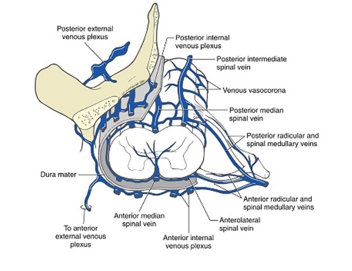
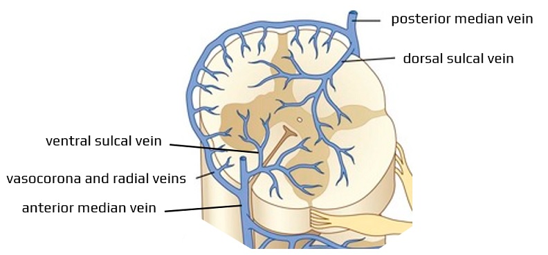
- small intramedullary veins drain into the longitudinal, tortuous venous plexus that forms along the entire length of the spinal cord
- ventral sulcal veins drain the medial portion of the anterior horn, the anterior commissure, and the white matter of the anterior funiculus
- the dorsal central veins drain the gray commissure and the white matter near the posterior median sulcus
- the remaining areas of the spinal cord are also drained centrifugally by peripheral radial veins
- anastomotic veins connect the radial and sulcal veins
- this venous plexus is formed by:
- anterior spinal vein (which collects blood from the sulcal and radial veins) – usually paired in the cervical and thoracic regions and a single vessel in the lumbar segment
- posterior spinal vein and 2 lateral longitudinal veins that pass behind the nerve roots
- venous vasocorona (connecting longitudinal trunks)
- from both the anterior and posterior veins, blood is drained to the internal vertebral venous plexus (the Batson venous plexus) via several ventral and dorsal radiculomedullary veins
- radiculomedullary veins exit the dura, usually along the nerve root
- the largest radiculomedullary vein called the great anterior radiculomedullary vein (GARV), drains the thoracolumbar cord and may be mistaken for the artery of Adamkiewicz
- the internal vertebral plexus consists of two anterior longitudinal veins running on either side of the posterior longitudinal ligament and two posterior veins running anterior to the ligamentum flavum
- the internal vertebral plexus is connected to the external vertebral plexus via the intervertebral foramina (intervertebral veins)
- veins around the spine lack valves, allowing blood to flow in different directions depending on the pressure gradient
- the external vertebral plexus drains into the vertebral veins of the neck and the segmental veins of the trunk
- cervical cord → vertebral, deep cervical, and jugular veins
- thoracic cord → left hemiazygos and accessory hemiazygos veins and right azygos vein
- lumbar cord → azygos and ascending lumbar veins


