NEUROIMAGING

Neuroimaging During Pregnancy and Lactation

David Goldemund M.D.
Updated on 21/03/2024, published on 08/09/2023

  • ultrasound and magnetic resonance imaging (MRI) are not associated with increased risk and are the preferred imaging modalities for pregnant women (ACOG Guidelines 2017)
  • still, both methods should be used with caution (especially MRI) and only when there is a relevant clinical question that needs answering or when medical benefits to the patient can be expected
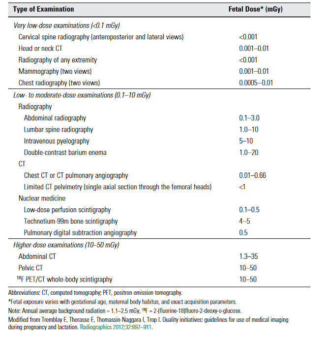
Fetal radiation dose caused by different diagnostic methods
Computed tomography (CT)
Content available only for logged-in subscribers (registration will be available soon)
Magnetic resonance imaging (MRI)

Exposure to non-contrast MRI

  • MRI is the preferred imaging modality in pregnancy, whether for maternal or fetal reasons.
  • using a 1.5 T scanner is recommended
  • to date, there are no known adverse effects or specific consequences for fetuses exposed to non-contrast MRI
    • published data indicate no increased risk of fetal malformations, even in the first trimester  [Ray, 2016]
    • there is a growing body of experience with fetal MRI (used to detect brain developmental defects, placental abnormalities, and more) [Griffiths, 2005]

Exposure to gadolinium-based contrast agent (GBCA)

Pregnancy

  • gadolinium contrast agents should be avoided (particularly during the period of organogenesis) unless essential to address a relevant clinical question
    • gadolinium crosses the placenta and has a significantly prolonged elimination from the fetal circulation with a risk of deposition in fetal tissues
    • animal studies indicate teratogenicity with high and repeated doses of IV gadolinium
    • analyze the risk-benefit; the patient must give informed consent after a discussion of risks and benefits
    • in vascular neurology, contrast administration can be avoided by using time-of-flight (TOF) sequences
    • if contrast agent administration is necessary, prefer macrocyclic GBCAs with the lowest feasible dose [Copel, 2017]

Lactation

  • there is a limited excretion of GBCAs into breast milk (< 0.04% in the first 24 hours, and the infant absorbs < 1%)  [Copel, 2017]
  • GBCAs should only be used when clinically necessary and non-postponable
  • there is no proven benefit of interrupting breastfeeding after GBCAs administration
Neurosonology
  • neurosonology is safe for both the mother and the fetus, and it is the preferred method for diagnosing and monitoring occlusions and stenoses during pregnancy
  • ultrasound is also a safe method for examination of the fetus, but the ALARA principle should be followed regarding the amount of energy used (max. intensity < 720 mW/cm2)
    • the thermal effect on the fetus is highest with the color and Doppler mode and lowest with the B mode
    • it’s advisable to use obstetric presets for these examinations
Angiography (DSA)
  • DSA is mostly reserved for interventional procedures, such as mechanical thrombectomy (MT), aneurysm coiling, etc.
    • pregnant women were excluded from randomized trials with MT, but the benefit can be assumed ⇒ pregnancy is, therefore, a relative contraindication to MT
    • published case reports have shown favorable maternal and fetal outcomes [Blythe, 2019]
  • careful fetal shielding with a lead apron is essential during such procedures

You cannot copy content of this page

Send this to a friend
Hi,
you may find this topic useful:

Neuroimaging during pregnancy and lactation
link: https://www.stroke-manual.com/neuroimaging-during-pregnancy-and-lactation/