STROKE CONSEQUENCES
Poststroke spasticity
Updated on 16/03/2024, published on 14/01/2024
- poststroke spasticity (PSS) is one of the symptoms associated with central (upper) motoneuron lesions
- studies suggest that ischemic lesions involving the basal ganglia, thalamus, insula, and white matter tracts especially the internal capsule, corona radiata, external capsule, and superior longitudinal fasciculus, are predictive of PSS
- it is characterized by increased muscle tone causing stiffness and difficulty with movement; along with residual paresis, it is a major contributor to functional disability → Consequences of stroke
- poststroke spasticity:
- limits self-care and causes problems with activities of daily living (ADL)
- may interfere with hygiene and nursing care
- causes complications such as pressure ulcers, discomfort and pain, or skin infections
- central pain, “frozen shoulder” pain, etc.
- may lead to contractures or subluxations that limit physical therapy
- a contracture is defined as a decreased elasticity of a muscle, tendon, ligament, joint capsule, and skin, leading to increased resistance to passive stretching, similar to spasticity. However, the difference between the two is that contractures do now show any velocity-dependent changes in movement or limb positioning
- it typically develops in the first three months after stroke, with a varying incidence (~ 35%; quite a wide range, however, is reported in the literature) (Payenok,2019) (Bochkezanian, 2018) (Malhotra, 2011)
- reported incidence and prevalence vary widely due to differences in the definition and clinical measurement of spasticity, as well as differences in stroke severity, location, and chronicity
- reported incidence and prevalence vary widely due to differences in the definition and clinical measurement of spasticity, as well as differences in stroke severity, location, and chronicity
- early and aggressive rehabilitation therapy is essential for maximizing recovery and improving functional outcomes; rehabilitation programs typically focus on strengthening the affected limb, reducing spasticity, and teaching compensatory strategies
Definition and etiopatogensis of spasticity
- spasticity is defined as impaired sensorimotor control manifested by intermittent or continuous involuntary muscle activation
- it belongs to positive signs of the upper motor neuron syndrome (UMNS)
- caused by lesions proximal to the alpha motor neuron, resulting in an imbalance between the excitatory and inhibitory inputs at the level of the spinal cord
- the loss of descending inhibitory influences from the brainstem and cortex leads to hypersensitivity of the reflex arc with increased muscle tone and hyperreflexia
- there is a typical increase in resistance during passive stretching of the muscle and this resistance increases as the speed of stretching increases
- symptoms of UMNS are divided into negative and positive:
- negative symptoms – weakness, slowed movement, loss of dexterity, and rapid fatigability
- positive symptoms – spasticity with tendon hyperreflexia or clonus, spastic dystonia, pathologic skin reflexes (Babinski sign), synkinesis, co-contractions
- pure involvement of the pyramidal pathway (corticospinal tract originating in area 4 of motor cortex) would lead to paresis without spasticity
- positive symptoms of UMN are mainly due to lesions of the so-called parapyramidal pathways (from area 6), which terminate at the bodies of interneurons in the anterior corners of the spinal grey matter
- in addition to stroke, many other conditions may lead to spasticity (such as cerebral palsy, anoxia, traumatic brain injury, spinal cord injury, multiple sclerosis, and other neurodegenerative diseases, etc.)
Classification
- according to localization of the lesion
- cerebral (cortical, subcortical, brainstem lesion)
- spinal
- according to the local distribution of spasticity into:
- focal
- multifocal
- segmental
- hemispastic
- paraspastic
- generalized
- according to severity into:
- mild (increased tone, at most a slight restriction in range of motion, mild spasms or clonus)
- moderate (more pronounced hypertonus, greater restriction of range of motion, possibility of developing contractures, problems in releasing handshake, walking, and turning in bed)
- severe (marked hypertonus and marked limitation of range of motion, contractures, problems with moving, sitting)
- spasticity should be assessed using standardized validated clinical assessment scales such as the modified Ashworth scale (MAS)
Clinical presentation
- in some cases, the manifestations are subtle, and in others, muscle tone is increased to the point of joint immobilization
- spasticity is characterized by an increase in resistance during passive stretching of the muscle, and this resistance increases as the rate of stretching increases
- cerebral-type spasticity is usually focal or multifocal, with a greater proportion of extensor spasms, especially in the lower limbs
- there are fewer flexor spasms, and clonus is less pronounced
- other signs (both positive and negative) of central motoneuron lesion occur simultaneously (see table)
- paresis (palsy)
- the main negative manifestation of central motoneuron syndrome and the main clinical manifestation that the patient is aware of
- the impairment of muscle strength varies from mild paresis to plegia and is usually the main cause of the patient’s disability
- muscle strength and movement coordination are also weakened due to co-contractions and spastic dystonia
- hyperreflexia
- spastic dystonia, unlike spasticity, is present at rest when there is no free movement; it may sometimes decrease with prolonged passive stretching
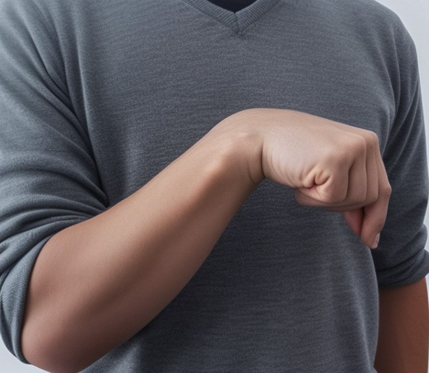
- PSS usually exhibits a focal or multifocal pattern, with flexion-pronation type observed in the upper extremities and extension type in the lower extremities ( Wernicke-Mann hemiparesis – the hand and fingers are held adducted with the forearm flexed, the leg is extended with plantar flexion of the foot so that it moves in a curved fashion)
- co-contraction
- synkinesis, etc.
- paresis (palsy)
| negative signs |
positive signs |
|
|
| Upper Limb | arm adduction arm intrarotation elbow flexion forearm pronation hand flexion thumb in palm clenched fist |
| Lower Limb | thigh adduction hip flexion knee flexion knee extension leg extension foot inversion toe extension finger flexion |
These movements should be tested and scored
- muscle hypertonus assessment – modified Ashworth Scale
- range of motion testing (goniometry)
- functional independence test
- functional assessment tested by an occupational therapist (Instrumental ADL)
- pain assessment – Visual analogue scale (VAS)
- VAS consists of a 10 cm line, with two end points representing 0 (‘no pain’) and 10 (‘pain as bad as it could possibly be’)
- Disability Assessment Score (DAS)
- quality of life assessment, e.g., the SF-36 Short Form Quality of Life Questionnaire
The modified Ashworth scale is the most universally accepted clinical tool used to measure spasticity. While popular, it has been criticized because of its inability to differentiate between the many factors that can contribute to resistance to passive stretch. Other methods have been proposed, including the Modified Tardieu Scale, Wartenberg Pendulum Test, Clinical Gait Analysis, Penn Spasm Frequency Scale, Visual Analog Scale, and Spinal Cord Assessment Tool for Spasticity
|
Modified Ashworth Spasticity Scale
|
|
| 0 |
no increase in muscle tone
|
| 1 | slight increase in muscle tone, with a catch and release or minimal resistance at the end of the range of motion when an affected part(s) is moved in flexion or extension |
| 1+ | slight increase in muscle tone, manifested as a catch, followed by minimal resistance through the remainder (less than half) of the range of motion |
| 2 | a marked increase in muscle tone throughout most of the range of motion, but affected part(s) are still easily moved |
| 3 | considerable increase in muscle tone, passive movement is difficult |
| 4 | affected part(s) rigid in flexion or extension |
[Bohannon a Smith, 1986]
Management
- treatment of patients with poststroke spasticity must be individualized and requires a multidisciplinary approach (neurologist, doctor of physiotherapy, orthopedist, physiotherapist, caregiver)
- a comprehensive initial assessment and the establishment of realistic treatment goals are necessary
- an important goal is improving the hand function – grasping, holding, and manipulating objects
- in the lower extremities, some degree of spasticity may be beneficial, as it allows stable standing even in severe paresis
Physiotherapy
- rehabilitation and occupational therapy is recommended by a rehabilitation doctor, who further monitors the progress of the rehabilitation process
- individualized rehabilitation should start as soon as possible, from the first day, with a multidisciplinary rehabilitation team
- the goals of physiotherapy are to actively participate in restoring trunk and limb mobility, including gait training, preventing contractures, maintaining joint mobility, and physiological limb length
- orthotic devices can be used to support the proper functional position of the limbs
- occupational therapy begins simultaneously with physiotherapy. Occupational therapists work with physiotherapists and patients to create a plan for specific fine motor skill training based on sensory-motor functional therapy. They also assess and train self-sufficiency in activities such as mobility in bed, transfers, verticalization, dressing, personal hygiene, orientation, communication, and cooperation.
Botulinum neurotoxin (BoNT)
- first choice for focal/multifocal poststroke spasticity in combination with neurorehabilitation
- the efficacy and safety of BoNT in the treatment of upper limb spasticity has been demonstrated in a series of randomized, double-blind, placebo-controlled clinical trials
- the therapy reduces muscle tone and pain, prevents the development of deformities, improves activities of daily living (dressing, hygiene), and reduces caregiver burden
- in many patients, therapy leads to an improvement in spastic limb function
- BoNT therapy should be initiated when spasticity becomes symptomatic – leads to painful conditions, worsens disability, or is an obstacle to physiotherapy or occupational therapy
- some authors recommend treating developing spasticity within three months after the stroke to minimize or even prevent the development of significant spasticity with complications
- botulinum neurotoxins (BoNTs) act at the neuromuscular junction by inhibiting the release of acetylcholine, a neurotransmitter vital for muscle contraction
- BoNT binds with high affinity to presynaptic neurons
- the toxin is then internalized into the cell by the mechanism of endocytosis
- once inside, the toxin cleaves specific SNARE proteins (e.g., SNAP-25, VAMP/synaptobrevin) which disrupts the SNARE-mediated fusion of acetylcholine-containing vesicles with the presynaptic membrane
- as a result, acetylcholine release is inhibited, leading to reduced neurotransmission at the neuromuscular junction
- nevertheless, the axon is able to regenerate SNARE proteins, which allows for the resumption of neurotransmitter (acetylcholine) release into the synaptic cleft
- consequently, neuromuscular transmission is restored, leading to the recovery of muscle function
- the clinical effect of BoNT application occurs after a few days, with a maximum effect after 3-4 weeks, and subsides in 3-4 months. Therefore, most patients require repeated application of BoNT, with at least a three-month interval between sessions recommended
Inclusion criteria:
- moderate to severe spasticity lasting > 2 months
- focal distribution of spasticity
- no further improvement after rehabilitation treatment
- participation in an active rehabilitation and treatment program
Exclusion criteria:
- fixed contractures and joint deformities
- pregnancy
- neuromuscular diseases
- anticoagulant treatment
Other local therapies
Intrathecal application of baclofen
- this treatment is considered a very effective method for reducing muscle hypertonus, reducing the frequency of painful muscle spasms and improving the overall quality of life
Neurosurgical and orthopedic treatment
- DREZ (Dorsal Root Entry Zone) interruption
- lesion of the afferent fibers of the posterior spinal root entry zone in patients with focal spasticity of the leg and, less commonly, of the hand accompanied by chronic pain
- this indication is very rare in PSS
- hand or leg tendon transfer
Monitoring of therapy
- BoNT therapy is part of comprehensive care for patients with poststroke spasticity
- the effect of therapy should be regularly evaluated


