ISCHEMIC STROKE / CLASSIFICATION AND ETIOPATOGENESIS
Behçet disease
Updated on 27/06/2024, published on 16/02/2024
- Behçet disease is characterized by the inflammation of blood vessels (vasculitis) of multiple organ systems
- etiology remains unclear; genetic predisposition (HLA-B51), environmental factors, and immune system dysregulation are thought to be involved
- etiology remains unclear; genetic predisposition (HLA-B51), environmental factors, and immune system dysregulation are thought to be involved
- usual onset between 20-30 years of age, more common in males (2-5:1)
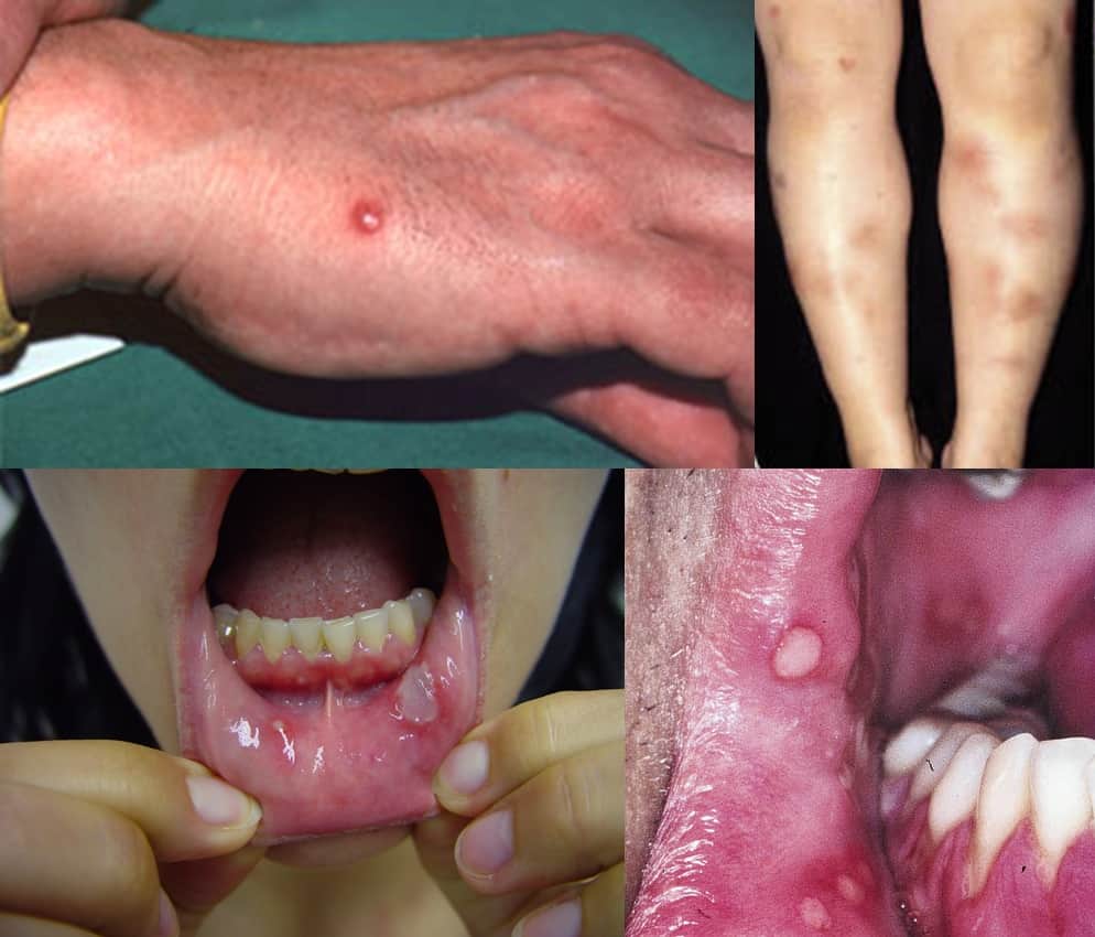
- characterized by recurrent ulceration in the mouth and external genitalia, as well as involvement of the eyes (uveitis), joints, skin, large vessels, and the nervous system
- the disease is more common in the Mediterranean, Middle East, and East Asia, with the highest incidence reported in Turkey; it is rarely reported in Europe
Clinical presentation
General symptoms
- recurrent uveitis (anterior and posterior) and keratitis leading to blindness
- oral and genital ulcers (mainly affecting the scrotum)
- initial presentation in ~60% of cases
- initial presentation in ~60% of cases
- skin involvement (nodular erythema)
- joint involvement (>50%)
- arthritis affecting ankles, knees, wrists, and elbows
- lasts for a limited period of several weeks and rarely results in permanent joint damage
- gastrointestinal and genitourinary tracts – mucosal ulcers
- cardiovascular manifestations (5-30% of cases)
CNS involvement (neuro-Behçet)
Parenchymal involvement
- meningoencephalitis, typically involving the brainstem
- occurrence up to one month after cutaneous manifestation
- remittent course, acute or gradual onset
- may clinically mimic MS
- absence of OCB, MS-atypical lesions on MRI
- lesions characteristics:
- T1 – hypointense
- T2 – hyperintense with vasogenic edema
- T1C+ – patchy enhancement
- DWI – isointense or slightly hyperintense
- MRS – decreased NAA + elevated lipid and choline/creatine ratio
- lesions localization:
- brainstem, BGG, and hypothalamus on T2 images
- do not correspond to vascular territories
- subcortical white matter is less commonly affected
- lesions often resolve after therapy
- atrophy is common in chronic course
- myelitis is less common (~ 10% of cases)
- myelitis can be the first manifestation of the disease; a history of recurrent uveitis or ulcerations can help to establish the correct diagnosis
- typical T2 hyperintense lesions extend over 2 segments on MRI
- pleocytosis is found in the cerebrospinal fluid
- increased erythrocyte sedimentation rate (ESR) indicates systemic inflammation
- myelitis can be the first manifestation of the disease; a history of recurrent uveitis or ulcerations can help to establish the correct diagnosis
- brainstem, BGG, and hypothalamus on T2 images
Vascular Behçet’s disease (less common)
- arterial infarctions (1-5%)
- Behçet’s disease is classified as a systemic vasculitis, although CNS vasculitis is rare
- involvement of large arteries is atypical for Behçet’s disease
- the role of microembolization due to a prothrombotic state is also considered
- aneurysms formation
- cerebral venous thrombosis (11-35%)
- non-specific symptoms can lead to delayed diagnosis
- usually involves multiple sinuses
Combination of parenchymal and vascular involvement
Diagnostic evaluation
| The International Criteria for Behçet’s Disease (ICBD) (Davatchi, 2014) |
||
|
Therapy
Anti-inflammatory drugs
- TNF-alpha inhibitors (e.g., infliximab, adalimumab) – used in refractory cases
treatment of joint manifestations and nodular erythema
Antithrombotics
- ASA 100 mg/d is sufficient for most stroke patients
- anticoagulants are used in combination with immunosuppressants to treat venous thrombosis
Prognosis
- the course of Behçet’s disease is variable, with periods of exacerbation and remission
- while it is a lifelong condition, many patients can achieve significant improvement in their symptoms and quality of life with appropriate management
- the overall activity often decreases over time


