ADD-ONS / ANATOMY
Collateral circulation assessment
Updated on 26/12/2023, published on 01/03/2022
- collateral cerebral circulation plays a critical role in maintaining blood flow to ischemic areas in the acute, subacute, or chronic phases after ischemic stroke
- good collateral circulation is associated with a favorable functional outcome and a lower risk of stroke recurrence
- in occlusions at the level of the circle of Willis and distally, leptomeningeal anastomoses (LMA) are mainly engaged
- the efficiency of collateral cerebral circulation can be assessed by evaluating perfusion distal to the occlusion/stenosis
- in acute stroke (⇒ estimation of prognosis and usefulness of thrombectomy, especially when CTP is not available)
- in the management of chronic extracranial steno-occlusive diseases (⇒ may help decide whether to perform revascularization)
- imaging methods used to assess cerebral collateral circulation:
- digital subtraction angiography (DSA)
- gold standard; currently used as a part of endovascular procedure; evaluation by the ASITN/SIR collateral scale helps predict the risk and benefit of acute endovascular treatment
- CT angiography
- traditional single-phase CTA (more reliable than MRA)
- CTA source image
- multiphase CTA (dynamic CTA)
- traditional single-phase CTA (more reliable than MRA)
- MR angiography
- time-of-flight MRA (TOF-MRA)
- phase-contrast MRA
- quantitative MRA (QMRA)
- CT perfusion
- MR perfusion
- neurosonology
- digital subtraction angiography (DSA)
- DSA is considered a gold standard; however, noninvasive imaging modalities are more commonly used
- there is no general agreement on the optimal collateral grading system based on noninvasive imaging modalities; further research is needed
| Imaging methods to assess the structure of the cerebral collateral circulation | Imaging methods to assess the function of the cerebral collateral circulation |
|
|
Collateral circulation assessment on DSA
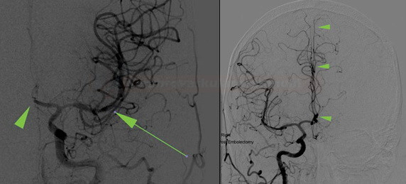
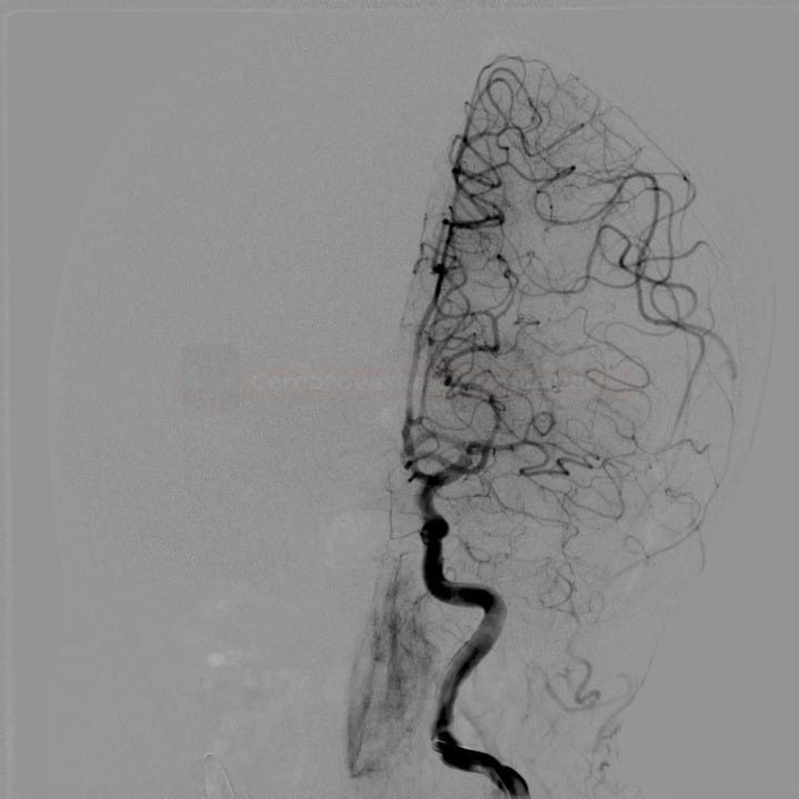
- DSA allows the assessment of collateral circulation dynamics; disadvantages are invasiveness and the need to examine all 4 main arteries
- it can be used during the endovascular procedure, e.g., to assess the collateral flow through the ACA in the case of M1 occlusion
- it is also necessary to assess the venous phase
|
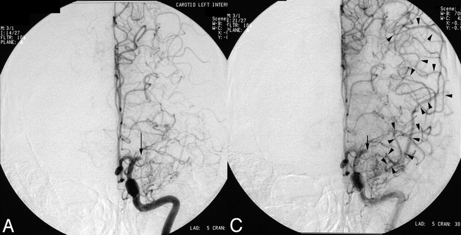
The American Society of Interventional and Therapeutic Neuroradiology/Society of Interventional Radiology (ASITN/SIR) grading system [Higashida,2003]
|
| Grade | Angiographic collaterals |
| 0 | no collaterals visible in the ischemic area |
| 1 | slow collaterals to the periphery of the ischemic area with persisting defect |
| 2 | fast collaterals to the periphery of the ischemic area with some persisting defect |
| 3 | collaterals with slow but complete filling of the ischemic area in the late venous phase |
| 4 | complete and rapid blood flow to the entire ischemic territory by retrograde perfusion |
|
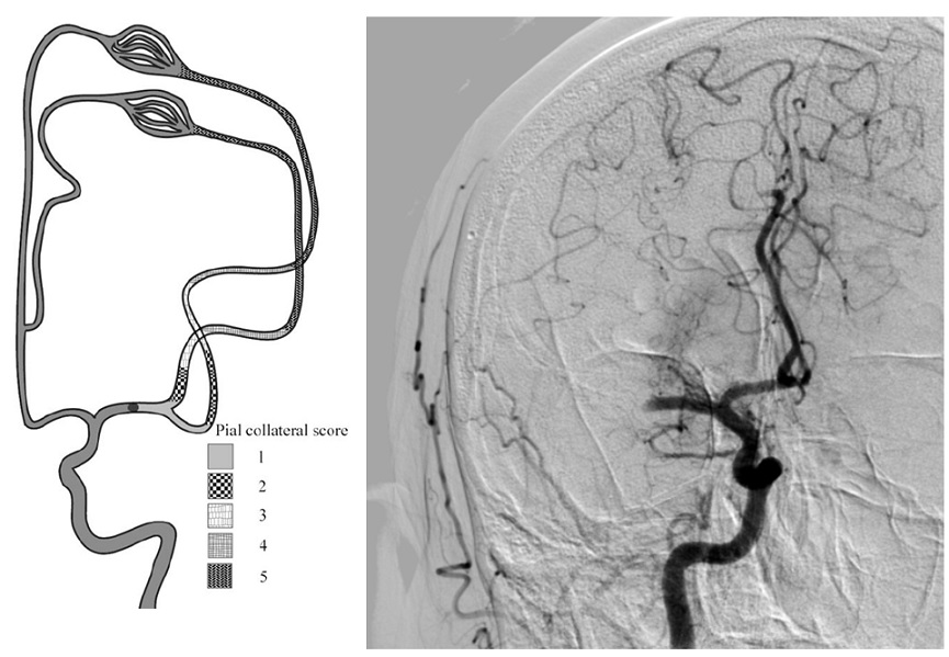
Pial Collateral Score [Christoforidis et al,2005]
|
- another DSA-based collateral grading system, that is less frequently used in clinical practice
- grade 1 – collaterals reconstitute the entire distal portion of the occluded vessel segment
- grade 2 – collaterals reconstitute vessels in the proximal portion of the segment adjacent to the occluded vessel
- grade 3 – collaterals reconstitute vessels in the distal portion of the segment adjacent to the occluded vessel
- grade 4 – collaterals reconstitute vessels two segments distal to the occluded vessel
- grade 5 – little or no significant reconstitution of the territory of the occluded vessel
- good collateral status (grades 1 and 2) has been correlated with smaller infarct volume, lower risk of hemorrhagic transformation, and lower modified Rankin Scale (mRS) at discharge
Collateral circulation assessment on CTA
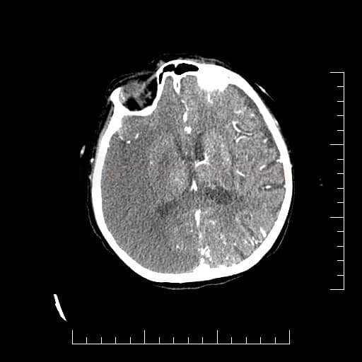
- in addition to the detection of occlusions, CTA also enables the analysis of collateral circulation; the presence of good collateral circulation correlates with smaller infarct size and predicts a better clinical outcome during reperfusion therapy
- a simple Collateral Score (CS) may be used for evaluation
- a semi-quantitative rapid comparison of collateral filling in the territory of the occluded artery compared to the contralateral hemisphere
- a single-phase and multiphase CTA (mCTA) can be used
- a limitation of conventional (single-phase) CTA is its static presentation; it is acquired during a short interval in the arterial phase, which can lead to an underestimation of delayed collateral circulation
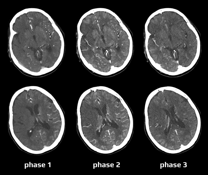
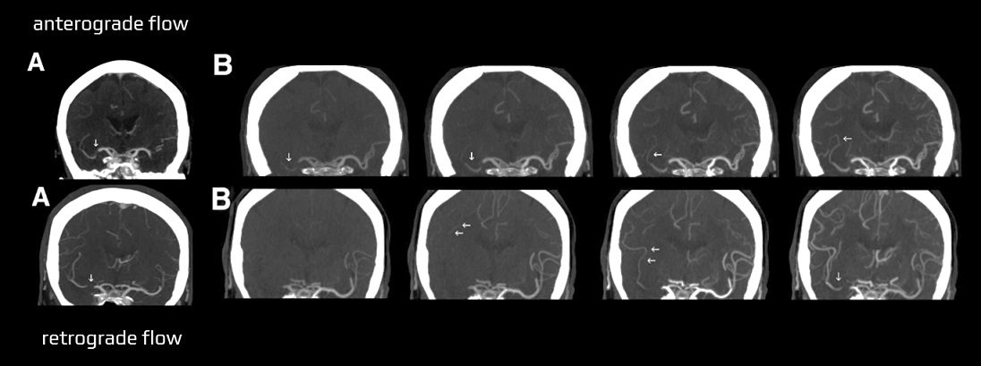
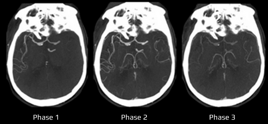
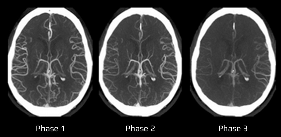
- dynamic information is provided by multiphase CTA (MP-CTA / mCTA)
- a total of 3-4 phases of intracranial CTA are performed using a reduced X-ray dose
- mCTA can differentiate between the absence of collaterals and delayed filling [Yang, 2008]
- mCTA can distinguish between minimal anterograde flow and retrograde collateral flow [Fröhlich, 2012]
- a total of 3-4 phases of intracranial CTA are performed using a reduced X-ray dose
The evaluation of CTA source images (CTA-SI) includes the following steps:
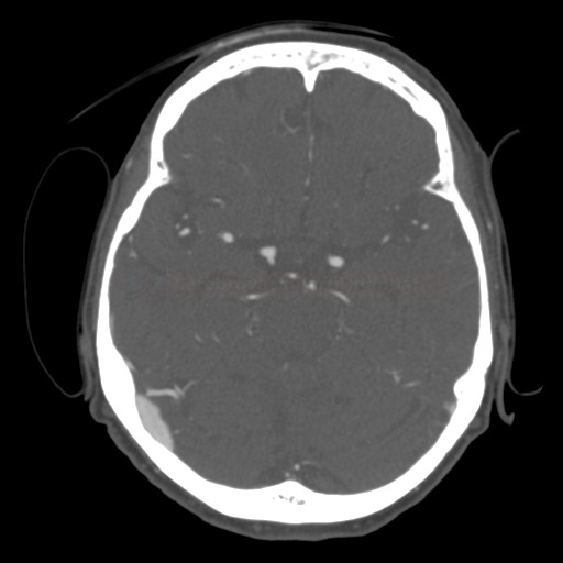
- check the circle of Willis for the presence and quality of communicating arteries, hypo/aplasia, etc.
- identify arterial occlusions and try to estimate their extent (thrombus length ⇒ Clot Burden Score (CBS)
- compare the filling of the arterial branches in both hemispheres
- evaluate the degree of the retrograde filling (optimally, the contrast agent should reach the distal end of the thrombus)
Collateral score in the anterior circulation (typically MCA)
| Miteff collateral grading on single-phase CTA (Miteff, 2009) | |||
| good | major MCA branches are reconstituted distal to the occlusion | ||
| moderate | some MCA branches are shown in the Sylvian fissure | ||
| poor | only the distal superficial MCA branches are reconstituted | ||
| Collateral status is graded in maximum intensity projection reconstructions (MIP) of single-phase CTA in axial, coronal, and sagittal planes in patients with MCA occlusion | |||
| Collateral Score (CS) on single-phase CTA [Tan, 2009] Based on single-phase CTA in patients with unilateral anterior circulation infarct |
|||
| Score | collaterals on CTA | ||
| 0 | absent collateral supply to the occluded MCA territory | ||
| 1 | collateral supply filling ≤50% but >0% of the occluded MCA territory | ||
| 2 | collateral supply filling >50% but <100% of the occluded MCA territory | ||
| 3 | 100% collateral supply of the occluded MCA territory | ||
| Higher grades are associated with better CT perfusion parameters (MTT, CBF, and CBV), smaller final infarct volume, smaller thrombus extent, and improved outcome | |||
| Collateral Score (CS) on multiphase CTA [Menon, 2015] |
|||
| Score | Collaterals on CTA |
||
| 0 | no vessels are visible in the affected hemisphere in any phase | ||
| 1 | only a few vessels are visible in the affected hemisphere in any phase | ||
| 2 | a filling delay of two phases in the affected hemisphere with a significantly reduced number of vessels in the ischemic territory, or one phase delay showing regions with no visible vessels | ||
| 3 | a filling delay of two phases in the affected hemisphere or a delay of one phase with a significantly reduced number of vessels in the ischemic territory | ||
| 4 |
a filling delay of one phase in the affected hemisphere, but the extent and prominence of pial vessels are the same | ||
| 5 |
no filling delay compared to the asymptomatic contralateral hemisphere, normal pial vessels in the affected hemisphere | ||
| A score of ≤ 3 indicates a poor prognosis | |||
Case series of mCTA can be seen here
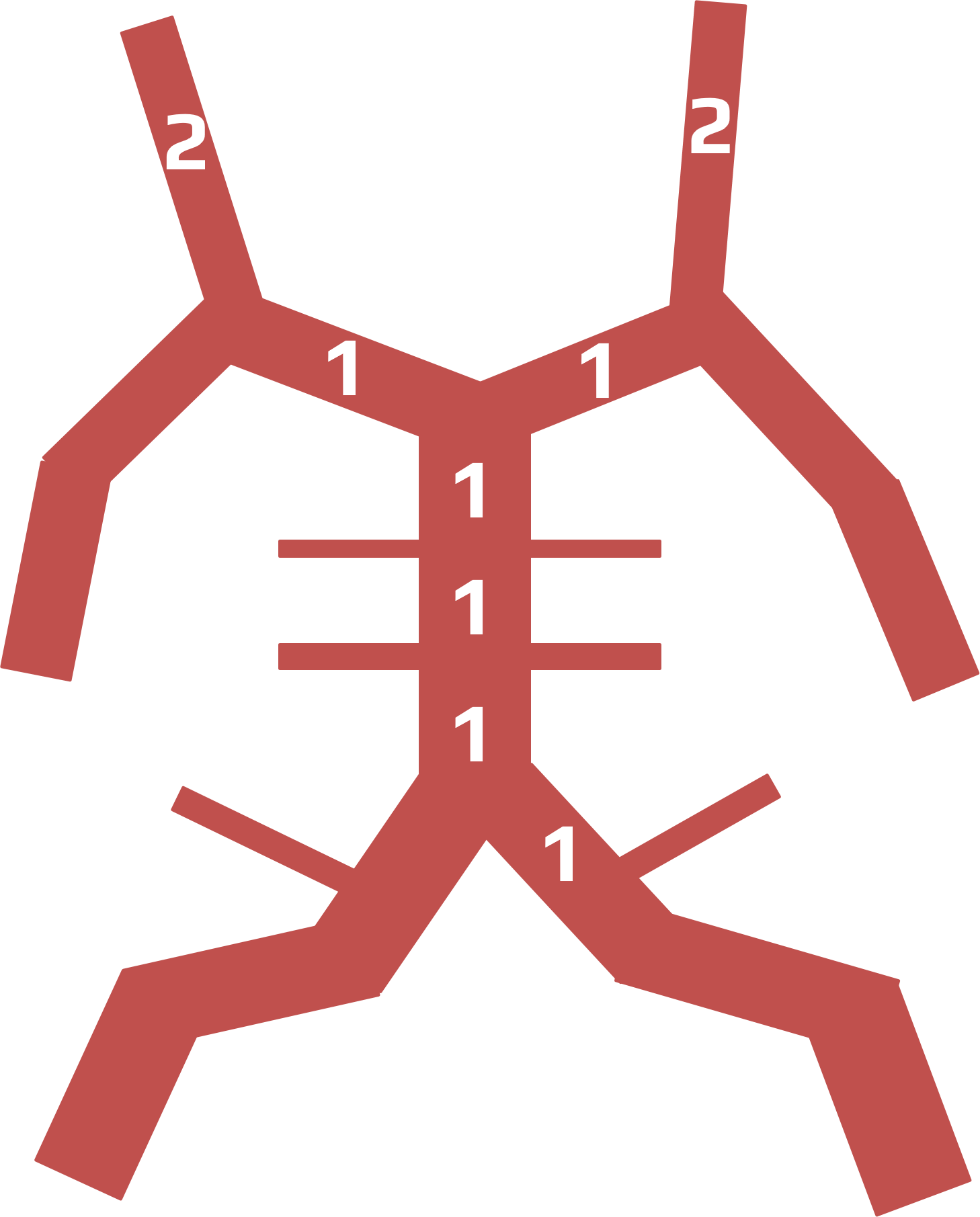
Basilar Artery on Computed Tomography Angiography (BATMAN) score
- the BATMAN score is a 10-point CTA–based grading system that incorporates thrombus burden and the presence of collaterals
- the posterior circulation is divided into 6 segments
- vertebral arteries (VA) – considered as 1 segment = 1 point
- posterior cerebral artery (PCA) – scored separately, 1 point each
- posterior communicant artery (PComA) – scored separately, 2 points each (or 3 points for fetal PCA)
- 3 segments of the basilar artery (BA) – 1 point each
- vertebral arteries (VA) – considered as 1 segment = 1 point
- patients with a lower BATMAN score were more likely to have a poor outcome – the absence of PComA (bilateral or unilateral) was the strongest predictor of poor clinical outcome (OR of 6.8) [Alemseged, 2017]
Posterior circulation CTA score
- 0 – no posterior communicating artery (PComA)
- 1 – unilateral PComA
- 2 – bilateral PComA
- the presence of bilateral PComA on CTA was associated with more favorable outcomes in patients with BAO undergoing mechanical thrombectomy [Goyal, 2016]
Posterior Circulation Collateral Score (PC-CS)
- max. 10 points (normal findings)
- AICA, PICA, SCA – assign 1 point to each patent artery (assess bilaterally)
- PComA – assign 1 point if PComA is smaller than the P1 segment, 2 points if larger
- patients with higher scores have better prognosis [Goyal, 2016]
Time of flight MRA
- another noninvasive method commonly used to assess the structure of cerebral collateral circulation
- assessment of leptomeningeal collaterals is limited due to relatively low spatial resolution
- TOF-MRA is typically used to assess primary collaterals via the circle of Willis
- sensitivity and specificity can be increased by combining the TOF-MRA with TCD/TCCD
Neurosonology
- TCD/TCCD is a noninvasive, low-cost method reflecting real-time cerebral blood flow velocity, collateral status, and cerebrovascular reactivity
- accuracy is highly operator-dependent
- TCD/TCCD can asses (directly or indirectly):
- collateral flow through AComA, PComA, ophthalmic artery, and leptomeningeal arteries
- the flow diversion phenomenon – high-velocity and low-resistance flow in the ACA or PCA in the presence of the MCA occlusion/severe stenosis (implies leptomeningeal collateral anastomoses between the ACA/PCA and the distal MCA branches
- collateral flow through AComA, PComA, ophthalmic artery, and leptomeningeal arteries

