Management of subarachnoid hemorrhage

Aneurysm clipping, artifact from the clip on CT scan
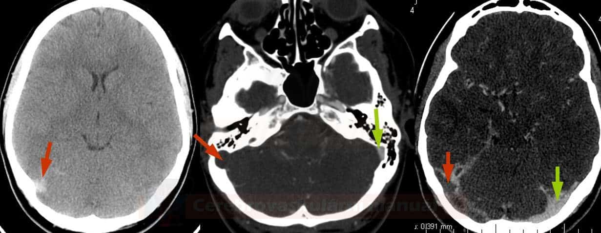
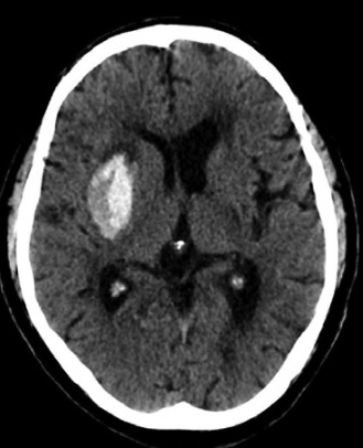
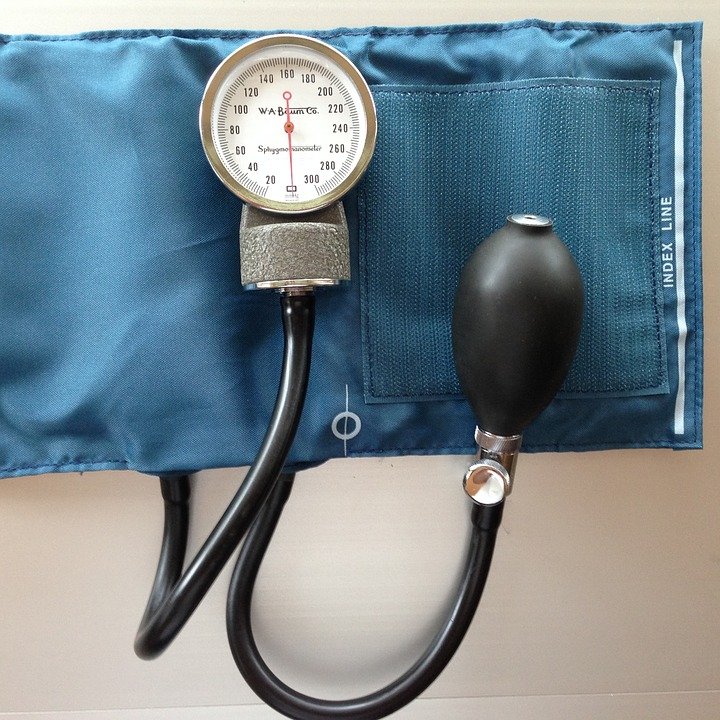
SUBARACHNOID HEMORRHAGE Management of subarachnoid hemorrhage David Goldemund M.D.Updated on 29/04/2024, published on 01/04/2023 [toc] Standard management of SAH focuses on: general care of acutely ill patients (incl. strict blood pressure control and maintenance of normovolemia) prevention and treatment of vasospasms detection and elimination of the aneurysm from the circulation prevention and management of complications  (intra- ... Read more

Cookie Policy

Cookie policy About this cookie policy This Cookie Policy explains what cookies are and how we use them, the types of cookies we use i.e, the information we collect using cookies and how that information is used, and how to control the cookie preferences. For further information on how we use, store, and keep your ... Read more

Management in the subacute phase of cerebral venous thrombosis

Hyperdense sign on NCCT (left), CT venography with defective contrast filling of the right transverse and sigmoid sinuses
CEREBRAL VENOUS SINUS THROMBOSIS Management in the subacute phase of cerebral venous thrombosis David Goldemund M.D.Updated on 15/05/2024, published on 08/03/2023 [toc] Anticoagulant therapy Drug choice acute parenteral therapy (usually using LMWH) is switched to oral anticoagulant therapy after 5-15 days (AHA guidelines, 2024) WARFARIN with a target INR 2-3  → see here DOACs  seem ... Read more

Blend Sign

Blend sign - a mix of hyperdense and relatively hypodense areas (difference > 18 HU) easily distinguishable by eye
Blend sign David Goldemund M.D.Updated on 21/04/2024, published on 25/02/2023 early hematoma growth is observed in approximately one-third of the patients with spontaneous intracerebral hemorrhage (ICH) the blend sign (BS) on baseline non-contrast computed tomography (NCCT) is a marker of increased risk of hemorrhage progression and a predictor of poor outcomes in patients with ICH  ... Read more

Dyslipidemia

Xanthelasma
ISCHEMIC STROKE / PREVENTION Dyslipidemia David Goldemund M.D.Updated on 12/04/2024, published on 20/02/2023 [toc] 2019 ESC/EAS Guidelines for the management of dyslipidaemias: lipid modification to reduce cardiovascular risk Definition of dyslipidemia the previously used term hyperlipidemia is now replaced by the broader term dyslipoproteinemia or dyslipidemia (DLP), which better reflects the fact that the pathology ... Read more

Arterial hypertension

Manual aneroid manometer
ISCHEMIC STROKE / PREVENTION / RISK FACTORS Arterial hypertension David Goldemund M.D.Updated on 21/11/2023, published on 20/02/2023 [toc] 2022 - WHO guideline for the pharmacological treatment of hypertension in adults 2018 - ESC/ESH Guidelines for the management of arterial hypertension Definition of hypertension hypertension is defined as office SBP values ≥ 140 mmHg and/or diastolic ... Read more

Diabetes mellitus

Diabetes mellitus
ISCHEMIC STROKE / PREVENTION / RISK FACTORS Diabetes mellitus David Goldemund M.D.Updated on 05/02/2024, published on 20/02/2023 [toc] Definition diabetes is a syndrome characterized by the inability of the body to process glucose and maintain its levels within physiological limits due to absolute (↓ secretion) or relative insulin deficiency (↑ resistance), resulting in altered glycide, ... Read more

Smoking

Smoking
ISCHEMIC STROKE / PREVENTION Smoking David Goldemund M.D.Updated on 07/05/2024, published on 20/02/2023 [toc] Smoking and risk of stroke smoking is classified as a disease (dg. F17 - drug addiction) cigarette smoking and passive smoking are both significant independent risk factors for ischemic stroke (1.5-2 fold risk)  [Peters, 2013] the excess risk of stroke is ... Read more

FAZEKAS scale

Fazekas
ADD-ONS / SCALES FAZEKAS scale David Goldemund M.D.Updated on 22/04/2024, published on 20/02/2023 a scoring system for simple assessment of white matter lesions (WML) on brain MRI scans was proposed by Fazekas in 1987 (Fazekas, 1987) WMLs often result from arteriolosclerosis (arteriolopathy)  the scale distinguishes two regions of white matter: Periventricular White Matter (PVWM) Deep ... Read more

Neurosonology in intensive care

Measurement of the third ventricle on the ultrasound
NEUROIMAGING / NEUROSONOLOGY Neurosonology in intensive care David Goldemund M.D.Updated on 25/01/2024, published on 15/02/2023 [toc] Monitoring of intracranial hypertension B-mode Midline shift transtemporal approach, using the thalamic plane set the depth to 15-16 cm from both temporal approaches, measure the distance of the midline structures (preferably the middle of the third ventricle) from the ... Read more

You cannot copy content of this page