INTRACEREBRAL HEMORRHAGE
Spot sign
David Goldemund M.D.
Updated on 20/12/2023, published on 13/04/2021
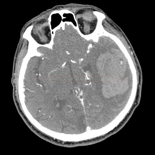
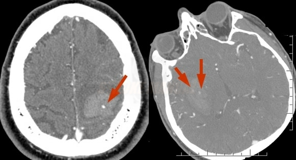
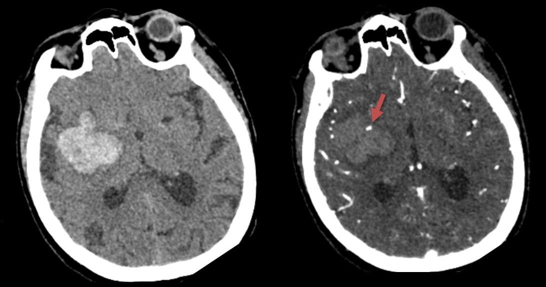
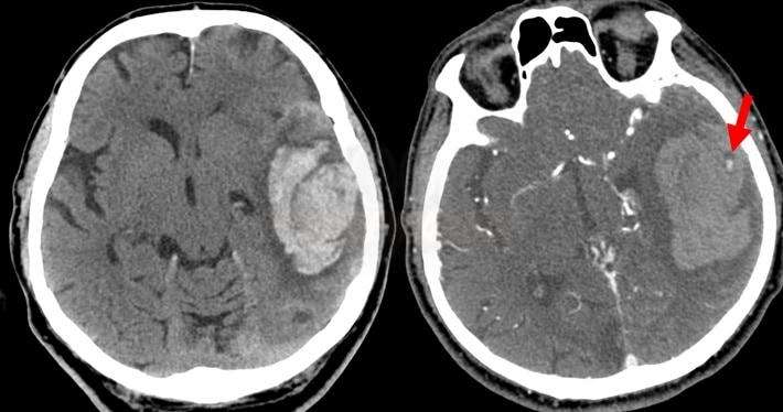
- the spot sign can be observed on post-contrast CT scans (typically CTA); it represents extravasation of contrast-enhanced blood within the hematoma
- the presence of the spot sign strongly correlates with early ICH growth (Wada, 2007)
- DDx of spot sign:
- AVM or aneurysm
- calcification (hyperdense on non-contrast CT scan)
- tumor
|
Definition of spot sign (extravasation on contrast-enhanced CT scan)
|
|
|
Spot sign score
- the spot sign score is a valuable predictor of early hematoma expansion, in-hospital mortality, and unfavorable outcome in patients with primary ICH
[Delgado Almandoz, 2009] [Havsteen, 2014]
- mortality rate based on spot sign score 2 ~ 30%, 3 ~ 50-70%, 4 ~ 70-90%, 5 ~ 100%
- the absence of a spot sign on the initial CT scan, followed by hematoma progression, can be explained by rebleeding or slow bleeding (detectable only on delayed post-contrast images)
|
Spot Sign Score (Delgado Almandoz)
|
|
|
Number of spot signs
1-2 ≥ 3
|
1
2
|
|
Maximum dimension
1-4 mm ≥ 5 mm |
0 1 |
|
Maximum density
20-179 HU
≥ 180 HU |
0 1 |


