Max-ICH score

ICH volume in mL (cm3) ≈ A (cm) x B x C /2. Volume is 4.4 mL

ADD-ONS / SCALES

Max-ICH score

David Goldemund M.D.
Updated on 29/08/2023, published on 08/06/2022

  • simple, reliable tool for predicting unfavorable long-term (12-month) functional outcome and mortality after intracerebral hemorrhage (ICH)
  • ICH involving both lobar and non-lobar regions should be scored based on the location where the ICH most likely originated
  • if 2 large ICHs occur simultaneously, more than 1 point referring to the ICH volume should be assigned
  • the maximum total score for a single hematoma (with/without IVH) is 9 points
  • each 1-point increase in the max-ICH score was associated with an OR of 1.24 for an unfavorable outcome  (Suo, 2018)
  • external validation comparing the ICH score and the max-ICH score shows a similar prognostic value (Schmidt, 2018)
Max-ICH score (0-9 points for a single ICH)
0-6
7-13
14-20
≥21
0
1
2
3
Age (years)
≤ 69
70-74
75-79
≥ 80
0
1
2
3
Lobar hematoma volume ≥ 30 mL
1
Non-lobar hematoma volume ≥ 10 mL
1
Intraventricular hemorrhage (IVH)
1
Oral anticoagulants
1
Max-ICH score and outcome (Suo, 2018)
Calculation of hematoma volume
volume in mL (cm3) ≈ A x B x C /2 (round or ellipsoid shape)
volume in mL (cm3) ≈ A x B x C /3 (irregular, separated, or multinodular shape) [Huttner, 2006]
A, B –  hemorrhage width and length – see image  (in cm)
C = height
  • number of CT slices showing hematoma x CT slice thickness
  • measure the height on sagittal reconstruction (2D tools in TOMOCON or other software)

For more precise measurement, count slices as follows:

  • slice with ≥75% area of hemorrhage counts as one slice
  • slice with 25-75% area of hemorrhage counts as 0.5 slices
  • slice with <25% counts as zero slices
ICH volume in mL (cm3) ≈ A (cm) x B x C /2. Volume is 4.4 mL

Black Hole Sign

Black hole sign on NCCT

Black Hole Sign

David Goldemund M.D.
Updated on 20/12/2023, published on 25/02/2022

  • the black hole sign (BHS) is a non-contrast CT scan marker of increased risk of early hematoma growth and a predictor of poor outcome in patients with intracerebral hemorrhage (Zhang, 2016)
    • BHS  is more common in patients with hematoma growth (31.9%) than those with no hematoma progression (5.8%) [Li, 2016]
  • it correlates well with spot sign, which seems to be more reliable and sensitive [Sporns, 2017]
Black hole sign on NCCT

Definition of Black Hole Sign [Li, 2016]
  • relatively hypoattenuating area (black hole) encapsulated within the hematoma (difference > 28 HU)
  • round, oval, or rod-like in shape
  • not connected with the adjacent brain tissue
  • the area should have a well-identifiable border

Capillary telangiectasia

Capillary telangiectasia on MR GRE and more sensitive SWI
INTRACEREBRAL HEMORRHAGE / VASCULAR MALFORMATIONS

Capillary telangiectasia

David Goldemund M.D.
Updated on 06/11/2023, published on 24/02/2022

[toc]

  • the second most common vascular malformation after venous angioma (DVA) (16-20% of all malformations)
  • the lesion is composed of vessels resembling dilated capillaries (lumen 20-500μm) separated by normal brain tissue (unlike cavernous malformations)
  • approx. 2/3 of the lesions have a visible small draining vein
  • typical locations:
    • pons
    • cerebellum
    • spinal cord
  • the vast majority of telangiectasias are asymptomatic (incidental finding on MRI)
    • low potential for hemorrhage unless multiple (e.g., in Rendu-Osler-Weber syndrome, where AVMs may also be present)  Skin and mucosal manifestations of Hereditary Hemorrhagic Telangiectasia (Rendu-Osler-Weber)
    • may cause focal neurological deficits

Diagnostic evaluation

  • MRI
    • small, usually solitary lesions without mass effect in typical locations (pons, cerebellum, and spinal cord)
    • T1: iso- to a hypointense lesion
    • T1C+: enhancing lesion, draining veins may be seen in large telangiectasias
    • T2: normal or only slightly hyperintense
    • GRE/SWI: hypointense (low signal caused by slowed flow, not hemorrhage) [Castillo, 2001]
  • CT – usually negative or shows a nonspecific small enhancing lesion
  • DSA – negative
Capillary telangiectasia on MR GRE and more sensitive SWI
Brainstem capillary telangiectasia on T2 (left) and T1C+ (right) sequences
Pontine capillary telangiectasia with brush-like pattern (T1C+)

Differential diagnosis

Management

  • usually conservative approach
    • the lesion is difficult to access
    • almost always asymptomatic
  • MRI follow-up is not required if the imaging is typical

Intracerebral hemorrhage scales and scores

Spot sign

ADD-ONS / SCALES

Intracerebral hemorrhage scales and scores

Created 26.05.2020, last update 08.06.2022

ICH score
Content available only for logged-in subscribers (registration will be available soon)
Max-ICH score
Content available only for logged-in subscribers (registration will be available soon)
Spot sign
Content available only for logged-in subscribers (registration will be available soon)
HAS-BLED
  • a tool to guide the decision to initiate anticoagulation in patients with Afib
  • always compare the risk for major bleeding (calculated by the HAS-BLED score) with the risk of thromboembolic events (calculated by the CHA2DS2-VASc score) ⇒  does the benefit of anticoagulation outweigh the risk of bleeding?
  • a study comparing HEMORR2HAGES, ATRIA, and HAS-BLED showed superior performance of the HAS-BLED score compared to the other two scores
HAS-BLED score
Hypertension
uncontrolled BP (SBP >160 mmHg)
1
Abnormal liver/renal function
renal disease – dialysis, transplant, Cr >2.26 mg/dL or >200 µmol/L
liver disease – cirrhosis or bilirubin >2x normal or AST/ALT/AP >3x normal
1
1
Stroke previous stroke
1
Bleeding
prior major bleeding or predisposition to bleeding
1
Labile INR unstable INR, time in therapeutic range <60% 1
Elderly age ≥ 65 years
1
Drugs/alcohol
medication predisposing to bleeding –  aspirin, clopidogrel, NSAIDs
heavy alcohol use
1
1
HAS-BLED score
Pisters et al. annual ICH risk
Lip et al. annual ICH risk
0 1.1% 0.9%
1 1% 3.4%
2 1.9% 4.1%
3 3.7% 5.8%
4 8.7% 8.9%
5 12.5% 9.1 %
Not enough data for higher scores; risk is most likely > 10%

A score ≥ 3 is associated with an increased risk of major bleeding.
Frequent monitoring, DOAC use, or alternatives to anticoagulation (such as
LAA occlusion) are recommended.

SMASH-U
Etiologic Classification of Intracerebral Hemorrhage – SMASH-U [Meretoja, 2012]
incidence mortality at 3 months
Structural lesions (cavernous malformation, AVM) 5% 4 %
Medication (warfarin, DOAC, antiplatelet therapy) 14% 54 %
Cerebral Amyloid Angiopathy (CAA)
20% 22 %
Systemic disease (liver, kidney disease, thrombocytopenia/thrombocytopathies) 5% 44 %
Hypertension 35% 33 %
Undetermined 21% 30%
ABC
  • in addition to clinical factors, the ABC-bleeding risk score also incorporates the biomarkers: high-sensitivity troponin T, growth differentiation factor–15, and hemoglobin

ABC-stroke score

ABC-stroke score

ABC-bleeding score

ABC-bleeding score
ORBIT
  • The ORBIT bleeding risk score has a superior predictive ability for major bleeding in AFib patients compared to the HAS-BLED and ATRIA risk scores. The ORBIT risk score may provide a simple, easy-to-remember tool to assist in clinical decision-making [O´Brian,  2015]  [Hilkens, 2017]
Older age ( >75 y) 1
Reduced hemoglobin/Hct/anemia  (men <13 g/dL and Hct < 40%, women < 12 g/dL and Hct < 36% ) 2
Bleeding 2
Insufficient kidney function (GFR < 60 mL/min/1.73 m2) 1
Treatment with antiplatelets 1
Maximum score 7
score 0–2 – low risk ~ 2.4% / y
score 3 –  medium risk ~ 4.7% / y
score ≥ 4 – high risk ~ 8.1% / y
Spetzler-Martin
  • The Spetzler-Martin arteriovenous malformation (AVM) grading system assigns points for various angiographic features to predict the risk of surgery
Spetzler-Martin AVM grading scale (grade I-V)
score
Nidus size – largest nidus diameter on angiography
  • small  (< 3 cm)
  • medium (3-6 cm)
  • large (> 6 cm)
1
2
3
The eloquence of the adjacent brain
  • non-eloquent
  • eloquent
    • sensory, motor, language, or visual cortex
    • internal capsule
    • hypothalamus / thalamus
    • cerebellar peduncles (superior, middle, or inferior) and cerebellar nuclei
    • brainstem
0
1

Venous drainage → Anatomy of veins and sinuses

  • superficial veins
  • deep cerebral veins
0
1
Spetzler-Martin grade 3
HEMORR2HAGES
  • the HEMORR2HAGES score is used to stratify patients’ risk of bleeding when using anticoagulation for atrial fibrillation (Afib) in conjunction with situation-specific risks
  • a  systemic review comparing the performance of HAS-BLED, ATRIA, and HEMORR2HAGES recommended HAS-BLED for assessing major bleeding risk in Afib patients
    • HEMORR2HAGES showed higher diagnostic accuracy but was considered more difficult to use due to its complexity
Hepatic/renal disease
1
Ethanol abuse
1
Malignancy history
1
Older (age >75 y) 1
Reduced platelet count or function, including aspirin therapy 1
Re-bleeding risk (history of prior bleeding) 2
Hypertension (uncontrolled)
1
Anemia (Hgb <13 g/dL for Men; Hgb <12 g/dL for Women)
1
Genetic factors (CYP 2C9 single-nucleotide polymorphisms)
1
Excessive fall risk 1
Stroke history
1
Total points 12
The annual risk of bleeding
Score 0 ~ 1.9 %/y
Score 1 ~ 2.5 %/y
Score 2 ~ 5.3 %/y
Score 3 ~ 8.4 %/y
Score 4 ~ 10.4 %/y
Score ≥ 5 ~ 12.3 %/y

Cerebral arteriovenous malformation (AVM)

Cerebral arteriovenous malformation (DSA)

INTRACEREBRAL HEMORRHAGE / VASCULAR MALFORMATIONS

Cerebral arteriovenous malformation (AVM)

David Goldemund M.D.
Updated on 29/02/2024, published on 23/04/2021

[toc]

  • brain AVMs are congenital developmental defects (present in about 1.4-4% of the population); they are likely to grow over time ( AVMs are diagnosed at a mean age of 31 years)
  • AVM consists of an abnormally enlarged feeding artery and draining veins, between which lies a conglomerate of A-V junctions closely associated with the brain parenchyma (nidus)
    • feeding arteries (one or more) are enlarged and tortuous; in 10-20% of cases, an aneurysm develops, which is often the source of bleeding/rebleeding
    • in the nidus, there are abnormal arteries and veins without capillaries (arteries are directly connected to veins)
    •  intranidal aneurysm is frequent (up to 50%)
    • draining veins (one or more) are tortuous, dilated, may cause mass effect, venous aneurysms (venous pouches) may also be seen
    •  if the transition between the feeding artery and the draining vein is direct (without nidus), it is called arteriovenous fistula (another type of cerebral vascular pathology)!
  • localization of AVM
    • supratentorial: ~85%  (2/3 superficial, 1/3 deep)
    • infratentorial: ~15%
  • AVMs are solitary in most cases (multiple in < 5% of cases)
    • multiple AVMs are present in hereditary hemorrhagic telangiectasia (Osler-Weber-Rendu syndrome) or Wyburn-Mason syndrome (craniofacial arteriovenous metameric syndrome)
  • AVMs are often discovered incidentally on CT/MRI scans performed for other reasons
  • parts of the AVM have a histologically abnormal structure, gliosis or calcification are found around the lesion
  • a clinical manifestation usually occurs between 20-40 years of age (in contrast to DAVF, which occurs between 40-70 years of age)
  • considering treatment options, it is essential to determine
    • number of feeding arteries
    • size of the nest
    • type of drainage (superficial or deep vein system)

Classification

  • nidus composition
    • compact (or glomerular) nidus: abnormal vessels without the interposition of normal brain tissue
    • diffuse (or proliferative) nidus: no well-formed nidus is present; brain tissue is interspersed within the anomalous vessels
  • The Spetzler-Martin arteriovenous malformation (AVM) grading system assigns points for various angiographic features to predict the risk of surgery
Spetzler-Martin AVM grading scale (grade I-V)
score
Nidus size – largest nidus diameter on angiography
  • small  (< 3 cm)
  • medium (3-6 cm)
  • large (> 6 cm)
1
2
3
The eloquence of the adjacent brain
  • non-eloquent
  • eloquent
    • sensory, motor, language, or visual cortex
    • internal capsule
    • hypothalamus / thalamus
    • cerebellar peduncles (superior, middle, or inferior) and cerebellar nuclei
    • brainstem
0
1

Venous drainage → Anatomy of veins and sinuses

  • superficial veins
  • deep cerebral veins
0
1
Spetzler-Martin grade 3
Content available only for logged-in subscribers (registration will be available soon)

Clinical presentation

  • incidental finding in asymptomatic patients (~ 15%)
  • intracerebral, intraventricular, and subarachnoid hemorrhage (40-65%)
    • risk of bleeding 1-4%/year, risk of rebleeding about 20% in the first year
    • risk factors:
      • previous bleeding
      • deep location and drainage
      • concomitant aneurysm in the nidus or feeding arteries
    • mortality is approx. 10% for the first bleeding and 20% for the third bleeding
  • epileptic seizures (20-25%)
  • headache (15%)
  • focal neurologic deficits
    • a consequence of bleeding, mass effect, or steal syndrome
  • pulsatile tinnitus

Diagnostic evaluation

Content available only for logged-in subscribers (registration will be available soon)

Differential diagnosis

  • other cerebral vascular malformations   Vascular malformations
    • cerebral proliferative angiopathy (CPA)   Cerebral proliferative angiopathy (CPA)   Proliferative angiopathy ("diffuse AVM")
      • absence of early, extensive venous drainage
      • involvement of an entire lobe or hemisphere as a diffuse network with normal neural tissue intermingled with the abnormal vessels
      • more common in females (2:1) and presents at a mean age of 20 years
      • the risk of neurologic deficits after the surgical or endovascular treatment is substantial
    • craniofacial arteriovenous metameric syndrome (CAMS)
    • developmental venous anomaly (DVA)
    • dural arteriovenous fistula (DAVF)
  • malignant tumor

Management

  • higher Spetzler-Martin grade ⇒ higher surgical risk   Risk of surgery and radiosurgery according to Spetzler-Martin grade
    • grades 1 and 2: surgery is safe (good outcome in 95-100% of patients)
    • grade 3 requires individual assessment
    • grades 4 and 5 are associated with a high perioperative risk due to potential damage to the draining venous system that may serve both the AVM and surrounding healthy tissue. The alternative here is conservative management (good outcome in ~35%)
  • other factors increasing the risk of bleeding:
    • aneurysm in the feeding arteries or nidus
    • venous stasis

Conservative therapy

Content available only for logged-in subscribers (registration will be available soon)

Surgery/endovascular treatment

Content available only for logged-in subscribers (registration will be available soon)

Dural arteriovenous fistula (DAVF)

Monitoring of the external carotid artery (ECA) during retroauricular compression of the feeding occipital artery
INTRACEREBRAL HEMORRHAGE / VASCULAR MALFORMATIONS

Dural arteriovenous fistula (DAVF)

David Goldemund M.D.
Updated on 29/02/2024, published on 21/04/2021

[toc]

  • dural arteriovenous fistula (DAVF) is characterized by abnormal connections (shunts) between an artery and a vein; DAVF typically presents with tinnitus, hemorrhage, or venous hypertension
  • DAVF accounts for ∼ 10-15% of vascular malformations and most commonly affects patients aged 40-60 years
  • most DAVFs are idiopathic; some patients have a history of previous craniotomy, head trauma, or dural sinus thrombosis  [Gandhi, 2012]

Pathology

  • DAVFs usually have multiple feeders
    • supratentorial
      • middle meningeal artery, superficial temporal artery (from ECA)
      • ethmoidal branches of the ophthalmic artery
    • cavernous sinus (ICA and/or ECA branches)  → Carotid-cavernous fistula (CCF)
    • posterior fossa
      • vertebral arteries
      • occipital artery (fom ECA)
  • most typical drainage paths:

Classification

Type I – anterograde drainage directly into dural venous sinuses/meningeal veins
Ia – 1 feeding artery
Ib – >1 feeding arteries
Type II – anterograde drainage into dural sinuses/meningeal veins + retrograde drainage into subarachnoid veins
Type III – predominantly retrograde drainage into cortical veins with their dilatation, no dural sinus or meningeal venous drainage
Borden classification of DAVF
Cognard [Cognard, 1995]
This classification provides valuable data for the determination of the risk with each dural AV fistula and enables decision-making about the appropriate therapy
Type I – only anterograde drain into dural sinuses, benign course
Type II 

  • IIa – simultaneous retrograde drainage into the dural sinus  (intracranial hypertension in 20% of cases)
  • IIb – simultaneous retrograde drainage into cortical veins  (bleeding in 10%)
  • IIa+b – simultaneous retrograde drainage into dural sinus + cortical veins
Type III, IV, V – no dural sinus drainage (high risk of bleeding – 40-65%)

  • III – direct cortical venous drainage without venous ectasia
  • IV – direct cortical venous drainage with venous ectasia
  • V – spinal venous drainage (⇒ high risk of progressive myelopathy)
Cognard classification of DAVFs

Clinical presentation

Clinical presentation is variable and depends on the location of the fistula and the pattern of venous drainage. Common symptoms include:

  • pulsatile tinnitus (typically with sigmoid and transverse sinus drainage)
  • symptoms of venous hypertension/congestion
    • headache and facial swelling
    • hemorrhagic venous infarction
    • spinal myelomalacia
    • cranial nerve palsies (such as abducens palsy with diplopia)
    • ocular (orbital) symptoms – conjunctival chemosis and swelling (carotid-cavernous fistula) – DDx of cavernous sinus thrombosis!
    • intracerebral or subarachnoid hemorrhage (high risk, especially with type II and III) [Li, 2015]
  • DAVF can be high-flow and consume a significant portion of ejection fraction (EF) => exertional dyspnea, left ventricular hypertrophy (LVH)

Diagnostic evaluation

  • CT/MRI
    • more useful for detecting complications (hemorrhage, venous infarction, edema)
    • insufficient for diagnosing DAVF itself – vascular imaging must be added (however, enlarged arterial feeders or dilated pial vessels in the subarachnoid space may sometimes be seen)
  • vascular imaging (CTA/MRA or DSA)
    • often, multiple feeders are present without an intervening nidus
    • dural sinuses are filled with contrast during the arterial phase
    • dilated and tortuous cortical veins in the subarachnoid space
    • DSA remains the best method to accurately assess feeding vessels and the presence and extent of retrograde venous drainage
  • neurosonology
    • Doppler can show decreased resistance (RI< 0.45) and increased flow velocity
    • not as sensitive as CTA/MRA or DSA for diagnosing DAVFs
DAVF fed from the occipital artery (ECA branch)
DAVF - accelerated flow and decreased peripheral resistance in the ECA
In a patient presenting with pulsatile tinnitus, ultrasound revealed increased flow in the external carotid artery (ECA) 
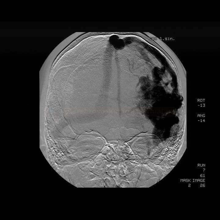
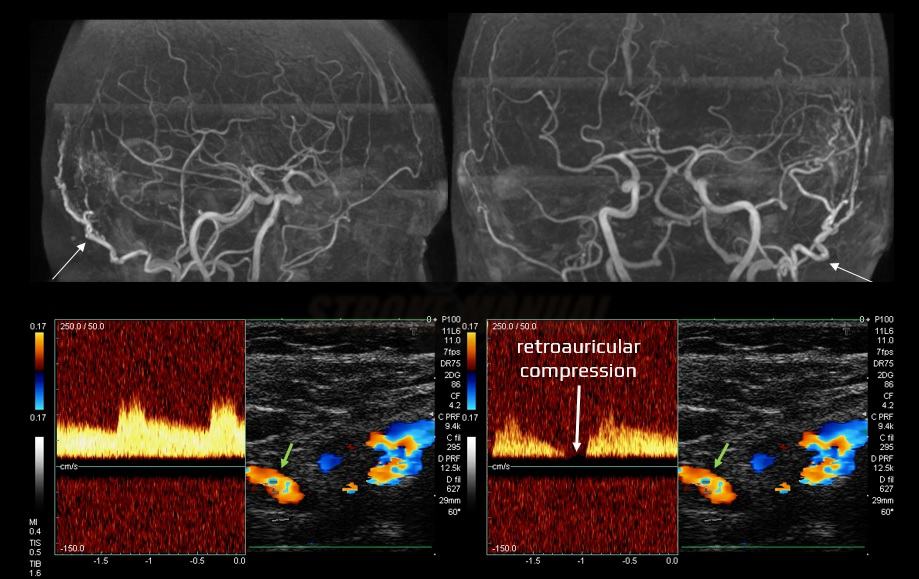
branch, characterized by aliasing and reduced pulsatility. Magnetic resonance angiography (MRA) subsequently confirmed the suspected diagnosis of a dural arteriovenous fistula (DAVF)
Dural arteriovenous fistula (DAVF) on DSA

Dural arteriovenous fistula (DAVF) on MRA

DSA reveals a fistula between the superficial temporal artery and the superior sagittal sinus. The white arrow indicates reflux from the sinus to the cortical veins

Management

  • treatment decision is based on the following factors:
    • type of the fistula
    • patient’s age and comorbidities (older patients and those with comorbidities may be better candidates for conservative treatment)
    • presence of symptoms attributable to the fistula

Conservative treatment

  • regular monitoring and lifestyle modifications are usually recommended for Borden I, Cognard I-IIa DAVFs
  • in mild cases, intermittent external compression of the feeding artery may be helpful (retroauricular compression in DAVF fed by the occipital artery)
Monitoring of the external carotid artery (ECA) during retroauricular compression of the feeding occipital artery

Endovascular and surgical treatment

  • typically considered for DAVFs with a higher risk of bleeding, such as Borden II and III or Cognard IIb-V DAVFs (an annual risk ~ 8%)
  • type I with severe tinnitus may also be indicated for endovascular treatment
Endovascular treatment
  • endovascular treatment involves occluding the abnormal connection between the artery and vein using embolization techniques
  • transarterial approach (TAE) – super-selective distal catheterization with injecting embolic agents into the feeding arteries
    • e.g., using Onyx [Lv, 2009]
  • transvenous approach (TVE) – involves occluding the venous drainage pathways; mainly used in cases with multiple small feeding arteries unsuitable for embolization
  • combined approach (TAE + TVE)
Surgery
  • typically reserved for cases where endovascular treatment is not possible or has been unsuccessful/partially successful
Stereotactic radiosurgery (SRS)
  • a minimally invasive second-line treatment option if endovascular or surgical treatment is not feasible or has been unsuccessful
  • typically dose of 20-30 Gy is used ⇒ irradiated vessels become thrombosed
  • obliteration is gradual, occuring within 2-3 years
  • unsuitable as primary treatment if there is reflux into cortical veins
Embolization of the direct arteriovenous fistula

Etiology and clinical presentation of intracerebral hemorrhage

Typical locations of bleeding in hypertensive patients
INTRACEREBRAL HEMORRHAGE

Etiology and clinical presentation of intracerebral hemorrhage

David Goldemund M.D.
Updated on 21/03/2024, published on 16/04/2021

[toc]

  • intracerebral hemorrhage (ICH) is characterized by spontaneous rupture of blood vessels within the cerebral parenchyma, leading to focal hematoma formation and subsequent mass effect
  • ICH accounts for approx. 10-20% of all strokes
  • the 30-day mortality is up to 40%, the annual mortality is ~50-60%, and severe deficits are common in survivors  [Broderick, 1993]
    • bleeding during antithrombotic therapy is associated with increased mortality (including DOACs and antiplatelets) – HR 1.3 on antiplatelet therapy, 1.4 on anticoagulation  [Apostolaki-Hansson]
  • ICH is a heterogeneous group in terms of etiology, clinical presentation, and therapy
  • a hematoma in the posterior fossa is always an acute, life-threatening condition
    • limited compliance can quickly lead to herniation upwards, transtentorially, or downward through the foramen magnum
Typical locations of bleeding in hypertensive patients

Intracranial vs. intracerebral hemorrhage

Intracranial hemorrhage refers to any bleeding within the skull, including the brain, its surrounding structures, and spaces. Intracerebral hemorrhage (ICH), a subset of intracranial hemorrhage, specifically denotes bleeding directly into the brain parenchyma.

Classification

Classification according to the etiology
  • primary (80%)
    • hypertensive arteriolopathy (70%)
    • amyloid angiopathy (CAA)
    • eclampsia
  • secondary (20%)
    • bleeding into a pre-existing lesion (tumor, ischemia)
    • coagulopathies (incl. drug-induced disorders)
    • malformations etc.
Classification according to ICH location
Supratentorial hemorrhage (85%)

  • cortical, lobar (30-35%)
  • deep hematomas – basal ganglia, internal capsule, and thalamus (55%)
Infratentorial hemorrhage (15%)

  • cerebellar (5-10%)
  • brainstem (5%)
Intraventricular hemorrhage (primary, secondary)
Intracerebral hemorrhage

Etiology

  • hypertensive arteriolopathy is the most common cause of intracerebral hemorrhage (ICH)
  • the relative risk of ICH in a patient with arterial hypertension compared to a person without hypertension is approximately 4
  • hypertension leads to bleeding by two mechanisms:
    • rupture of an artery affected by chronic hypertension
    • acute or subacute severe hypertension leading to rupture of a previously unaffected artery (malignant hypertension)
  • typical localization: basal ganglia, thalamus, cerebellum, pons   Typical localisations of hypertonic bleeding  Lenticulo-striate arteries
    • a secondary extension of the hematoma into the ventricles (hemocephalus) or subarachnoid space is possible
  • hypertension leads to hypertrophy and degeneration of the media of small arteries (lipohyalinosis, fibrinoid necrosis)
  • the findings that suggest hypertensive etiology:
    • history of hypertension
    • typical ICH localization
    • absence of any other apparent cause of bleeding
    • left ventricular hypertrophy
    • leukoaraiosis on CT/MRI   Hypertensive small vessel disease (microangiopathy)
    • hypertensive retinopathy
    • high blood pressure on admission is not a conclusive indicator of hypertensive disease; it may be a result of stress reaction and intracranial hypertension
  • aneurysm
    • 20-40% of SAHs have an IC hematoma component   Combination of SAH and ICH
    • rarely, rupture manifests as isolated ICH  [Li, 2016]
  • vascular malformations
MRI 0.25-0.7%
rebleeding 4.5%
MRI, DSA 0.2-0.4%
DSA, CTA 2-4 %
rebleeding 6-18%
MRI very low
DSA, MRA, CTA
type I – very low
type II, III – up to 8%
Carotido-cavernous fistula (CCF) DSA, MRA very low
Vascular malformations
  • anticoagulants (LMWHs, UFH, warfarin, DOACs)
    • the risk of ICH with long-term anticoagulant therapy is ~2% (risk is lower with DOACs)
    • risk of major bleeding with warfarin is 1.7% in those aged < 75 years, ~4.2% in those aged > 75 years; the risk of ICH is 0.6 vs. 1.8 (according to SPAF II)
    • hematomas are often non-homogeneous and multilobar (with a positive black hole and blend sign)
  • antiplatelet therapy
    • bleeding is more common with dual antiplatelet therapy (DAPT) and in combination with other risk factors
  • fibrinolytics
    • risk of symptomatic ICH (sICH) in acute stroke treated with tPA is ~ 6%
  • other coagulation disorders
    • leukemia, liver disease (related to alcoholism)
    • thrombocytopenia or thrombocytopathy
  • cerebral amyloid angiopathy (CAA)     Cerebral amyloid angiopathy
    • age >60 years or a positive family history
    • cognitive deterioration
    • β-amyloid deposits in small and medium-sized cerebral arteries
    • lobar hematomas, repeated/multiple hemorrhages (including microscopic ones)
    • leukoaraiosis on MRI and/or microbleeds on GRE    Cortical and subcortical CMBs
    • risk of recurrence ~10%/year (higher in individuals with APOE E2 and E4 positivity)
  • intracranial artery dissection (which is more likely to cause SAH or a combination of SAH+ICH)
  • vasculitis (polyarteritis nodosa, Wegener’s granulomatosis, SLE, Henoch-Schönlein, syphilis, primary CNS granulomatosis, etc.)
    • usually, hemorrhagic transformation of ischemia that must be distinguished from a primary hematoma
  • occasionally, it may be difficult to distinguish between a traumatic hematoma and a spontaneous hematoma that was the cause of the fall
  • signs of a traumatic etiology:
    • history of significant head trauma
    • CT scan with hemorrhagic contusions in the frontal and temporal lobes +/- concomitant SDH, EDH, or a traumatic SAH   Post-traumatic hemorrhagic contusions 
    • hemosinus Post-traumatic hemosinus on the CT scan
    • skull fracture visible in the bone window Bone fracture (the CT scan in the bone window)
  • cocaine, pseudoephedrine, amphetamine
  • drugs often cause lobar hematomas
Etiologic Classification of Intracerebral Hemorrhage – SMASH-U [Meretoja, 2012]
incidence mortality at 3 months
Structural lesions (cavernous malformation, AVM) 5% 4 %
Medication (warfarin, DOAC, antiplatelet therapy) 14% 54 %
Cerebral Amyloid Angiopathy (CAA)
20% 22 %
Systemic disease (liver, kidney disease, thrombocytopenia/thrombocytopathies) 5% 44 %
Hypertension 35% 33 %
Undetermined 21% 30%

Clinical presentation

Reliable clinical differentiation of cerebral ischemia from hemorrhage is not possible
  • sudden, apoplectic onset
  • personal medical history
    • dementia? (potential amyloid angiopathy)
    • hematologic disorder, antiplatelet, or anticoagulant therapy?
    • history of hypertension?
    • alcoholism, hepatopathy, renal disease?
    • history of bleeding or known malformations?
    • recent CEA or CAS? (risk of hyperperfusion injury)
  • focal neurologic symptoms (such as hemiparesis, aphasia, hemianopsia, etc., depending on hematoma location) →  Signs and symptoms of cerebral lesions
  • altered level of consciousness (up to 50%) – more common in ICH compared to ischemic stroke
    • the patient is usually somnolent or even soporous
    • initial coma occurs with extensive thalamic or brainstem hemorrhage, destructing the reticular formation
  • hypertension or hypertensive crisis (in up to 90%)
    • acutely decompensated chronic hypertension
    • stress-induced hypertension in otherwise normotensive patients
  • nausea and/or vomiting (24-50%)
  • headache (40%)
  • epileptic seizures (about 6%)
  • early improvement or fluctuation is not typical for ICH

Complications

Bleeding progression

  • about 1/3 of patients with ICH experience a 1/3 increase in hematoma within 3 hours of onset (2/3 of them within 1 hour) Asi u 1/3 pacientů s ICH dojde k nárůstu hematomu o 1/3 do 3 hodin (Kazui,1996)  [Kazui,1996]
    • perform a follow-up CT scan within 24 hours or immediately if the neurological status worsens
    • some protocols suggest performing a CT scan every 12 hours until the hematoma volume has stabilized
    • progression is most common in hematologic disorders but can also occur in typical hypertensive bleeding
  • progression of bleeding is associated with early neurological deterioration and poorer prognosis
  • radiologic predictors of progression:
  • hematoma progression in the following days is less typical and may indicate recurrent bleeding
    • particularly with hematologic disorders or vascular malformations (most commonly in aneurysms or AVMs)

Brain edema and intracranial hypertension

  • edema and intracranial hypertension develop shortly after the onset of bleeding, peaking between days 2 and 6
  • bleeding in the posterior fossa may lead to acute obstructive hydrocephalus

→ intracranial hypertension

Obstructive (non-communicating) hydrocephalus

  • the highest risk is associated with:
    • extensive intraventricular hemorrhage (primary or secondary)  Obstructive hydrocephalus following IVH
    • extensive cerebellar or brainstem hematomas causing direct compression of the cerebral aqueduct
  • ⇒ indication for acute surgery (EDV)

Epileptic seizures

Extracranial (systemic) complications

  • extracranial complications are similar to those observed in ischemic stroke → see here
  • some notes regarding blood pressure:
    • elevated blood pressure (BP) may result from decompensated chronic hypertension or be a stress reaction in previously normotensive individuals
    • elevated BP upon admission does not automatically imply a hypertensive etiology of the bleeding
    • normal BP upon admission increases the likelihood of bleeding from a vascular source  ⇒ perform vascular imaging (CTA, MRA, or DSA)

Prognosis

  • always make an initial rough estimation of the prognosis
    • 30-day mortality 35-50%, up to 70% for recurrent ICH
  • prognosis depends on the following:
    • age and general biological status (incl. comorbidities)
    • initial level of consciousness (LOC)
    • ICH location
    • hematoma size (ICH score)
      • GCS < 9 and ICH volume > 60ml ~ 90% mortality
      • GCS ≥ 9 and ICH volume < 30ml ~ 17% mortality
      • poor outcome is associated with an ICH score of 4-6
    • presence of spot sign, blend sign, and black hole sign
    • etiology of bleeding (SMASH-U)
    • acute phase complications (sepsis, ischemic stroke, prolonged mechanical ventilation, etc.)
  • functional recovery after ICH is highest in the first few weeks to months (greatest within 30 days)
    • early and long-term rehabilitation and ergotherapy are essential
    • for assessing the functional outcome, the Modified Rankin Scale (mRS) is frequently used
  • assess cognitive functions, as impairment is frequently observed among patients after ICH

Risk of recurrence

  • risk of ICH recurrence depends on etiology and risk factors
  • the estimated recurrence risk  is  1.2-7% per year across undifferentiated patients with ICH (with the highest event rate occurring in the first year after bleeding)
  • risk factors for the recurrence of ICH
    • advanced age
    • race (Black, Asian)
    • poorly controlled hypertension
    • history of prior ICH and ischemic stroke
    • ICH location (nonlobar<lobar)
    • etiology (increased risk with coagulation disorders, CCA, brainstem cavernous malformations, Moyamoya disease, AVM, tumors)
    • imaging features (lobar microbleeds, leukoencephalopathy, cortical superficial siderosis)
    • CHKD (can be a marker of atherosclerotic disease)
    • genetic features (carriers of apolipoprotein-E e2 or e4 genotypes)
ICH recurrence risk (annual)
Hypertonic bleeding (precise BP correction reduces RR by 50%) 1.1-4 %
Cerebral amyloid angiopathy (CAA)
7.5-20 %
AV malformation (AVM)
6-18 %
Cerebral cavernous malformation (CCM)
3.8-30%
Dural AV fistula 0.15 %

Follow up imaging

  • patients who fail to improve or deteriorate during the recovery phase need imaging to rule out recurrent bleeding
  • stabilized patients with hypertensive ICH don´t require additional imaging
    • features indicative of hypertensive bleeding: history of hypertension (HTN), subcortical microbleeds, no atypical imaging features, age ≥65 years
  • no definitively established etiology or baseline imaging features suggestive of an underlying cause require follow-up imaging performed after bleeding and edema have resolved
    • perform brain MRI  4-16 weeks after the ICH (incl. GRE/SWI and contrast-enhanced images);  if an underlying vascular cause is suspected, add noninvasive vascular imaging, such as CTA or MRA
    • MRI is optimal for detecting cerebral venous sinus thrombosis, vascular malformations, hemorrhagic transformation of an ischemic infarct, or neoplasms
    • contrast-enhanced CT of the brain is a reasonable alternative for those who are unable to undergo an MRI
  • stabilized CAA patients may not require additional imaging

Clinical features raising suspicion for an underlying cause

  • age <65 years
  • no history or new diagnosis of HTN
  • history of new-onset headaches
  • history of new-onset neurologic symptoms preceding ICH
  • thunderclap headache at the onset of hemorrhage
  • history of prior ICH (unless attributed to uncontrolled HTN or CAA)

Imaging features on baseline imaging raising suspicion for secondary cause of ICH

  • early perihematomal edema disproportional to the size of the hematoma
  • nonconfluent hemorrhage in the arterial vascular territory (probable ischemic infarction)
  • enhancement of intracranial vessels around ICH
  • multifocal hemorrhage
  • isolated intraventricular hemorrhage

ICH prevention

  • long-term precise blood pressure (BP) control is required in all ICH patients (AHA/ASA 2022 1/B-R)
    • this approach is supported by evidence from ischemic stroke prevention trials  (SPS3, RESPECT)
  • start therapy ASAP, combining pharmacological and nonpharmacological approaches
    • the first-choice drug is usually an ACE inhibitor; if not tolerated, use an angiotensin receptor blocker, thiazide diuretic, or calcium channel blocker
    • usually, a combination of drugs is necessary
  • acute BP management is discussed elsewhere (target 140-160 mmHg)
  • long-term BP should be maintained < 130/80 mmHg (AHA/ASA 2022 2a/B-NR)
  • aim for a BP < 120/80 mmHg in younger patients without major comorbidities (Teo, 2022)
  • a stepwise correction to achieve target values is suggested in the subacute phase (within 2 weeks)
  • outpatient monitoring is essential to ensure long-term proper BP control
  • reduced salt intake, healthy diet
  • cessation of smoking and excessive alcohol intake
  • treatment of sleep apnea, if present
  • regular physical activity
  • maintenance of a healthy body weight
  • avoidance of sympathomimetics
Content available only for logged-in subscribers (registration will be available soon)
  • epidemiological studies and clinical trials provide conflicting data
    • in patients with spontaneous ICH and an established indication for statin pharmacotherapy, the risks and benefits of statin therapy are uncertain (AHA/ASA guidelines 2022
    • the decision to use statins in patients with ICH depends on the individual assessment of the risk of ischemic events versus recurrent ICH
  • there is no strong evidence to discontinue the hypolipidemic therapy after ICH

    • a large 10-year nationwide cohort study from Taiwan found no association between statin dose and risk of recurrent ICH  (Tai, 201)
    • statins are associated with improved functional outcome and reduced mortality in patients with prior ICH  (Ziff, 2018)
    • data from the Danish registry show, that exposure to statins is not associated with an increased risk of recurrent ICH but was associated with a lower risk of any stroke (Gaist, 2023)
  • in patients with a high risk of hemorrhage (typically after recurrent parenchymal hematoma), a more cautious approach may be warranted, potentially involving alternative lipid-lowering drugs
  • in patients with spontaneous ICH, regular long-term use of nonsteroidal anti-inflammatory drugs (NSAIDs) is potentially harmful because of the increased risk of ICH (AHA/ASA 2022 3/B-NR)
  • prefer nonacetylated salicylates
  • the presence and extent of cerebral microbleeds and cortical superficial siderosis predict subsequent symptomatic ICH
  • incorporate available MRI results into decision-making regarding stroke prevention plans (avoid warfarin, apply strict BP management, etc.)  (AHA/ASA 2022 2b/C-LD)

Spot sign

Spot sign on CTA
INTRACEREBRAL HEMORRHAGE

Spot sign

David Goldemund M.D.
Updated on 20/12/2023, published on 13/04/2021

  • the spot sign can be observed on post-contrast CT scans (typically CTA); it represents extravasation of contrast-enhanced blood within the hematoma
  • the presence of the spot sign strongly correlates with early ICH growth (Wada, 2007)
  • DDx of spot sign:
    • AVM or aneurysm
    • calcification (hyperdense on non-contrast CT scan)
    • tumor
Definition of spot sign (extravasation on contrast-enhanced CT scan)
  • ≥ 1 contrast deposit inside the hematoma with increased density compared to the surrounding hematoma
  • density ≥ 120 HU
  • the lesion is not related to surrounding vascular structures
  • the lesion may be of any size and shape

Spot sign score

  • the spot sign score is a valuable predictor of early hematoma expansion, in-hospital mortality, and unfavorable outcome in patients with primary ICH Dependence of Spot Sign Score and outcome [Delgado, 2010] [Delgado Almandoz, 2009]   [Havsteen, 2014] 
    • mortality rate based on spot sign score 2 ~ 30%, 3 ~ 50-70%, 4 ~ 70-90%, 5 ~ 100%
  • the absence of a spot sign on the initial CT scan, followed by hematoma progression, can be explained by rebleeding or slow bleeding (detectable only on delayed post-contrast images)
Spot Sign Score (Delgado Almandoz)
Number of spot signs
1-2
≥ 3
1
2
Maximum dimension
1-4 mm
≥ 5 mm
0
1
Maximum density
20-179 HU
≥ 180 HU
0
1
Spot sign
Spot sign on baseline CTA
Parenchymal hematoma on NCCT (left), spot sign on CT angiography (right)
Spot sign on CTA

You cannot copy content of this page